42 nerves of the foot and ankle diagram
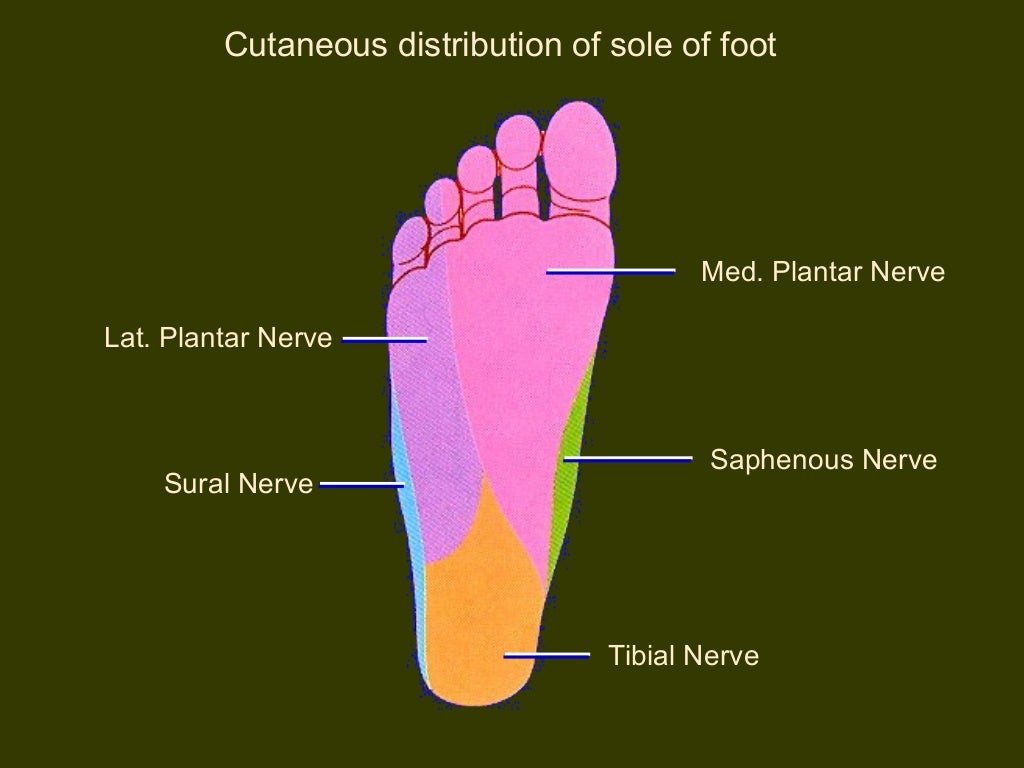
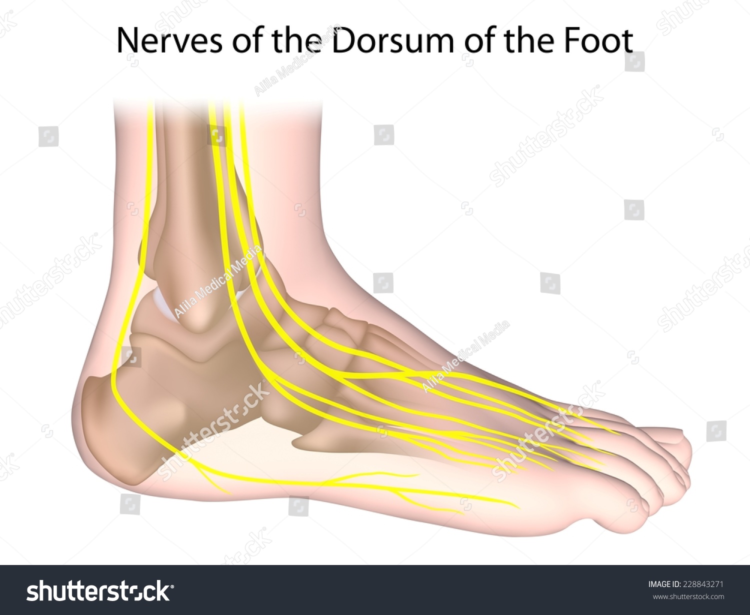
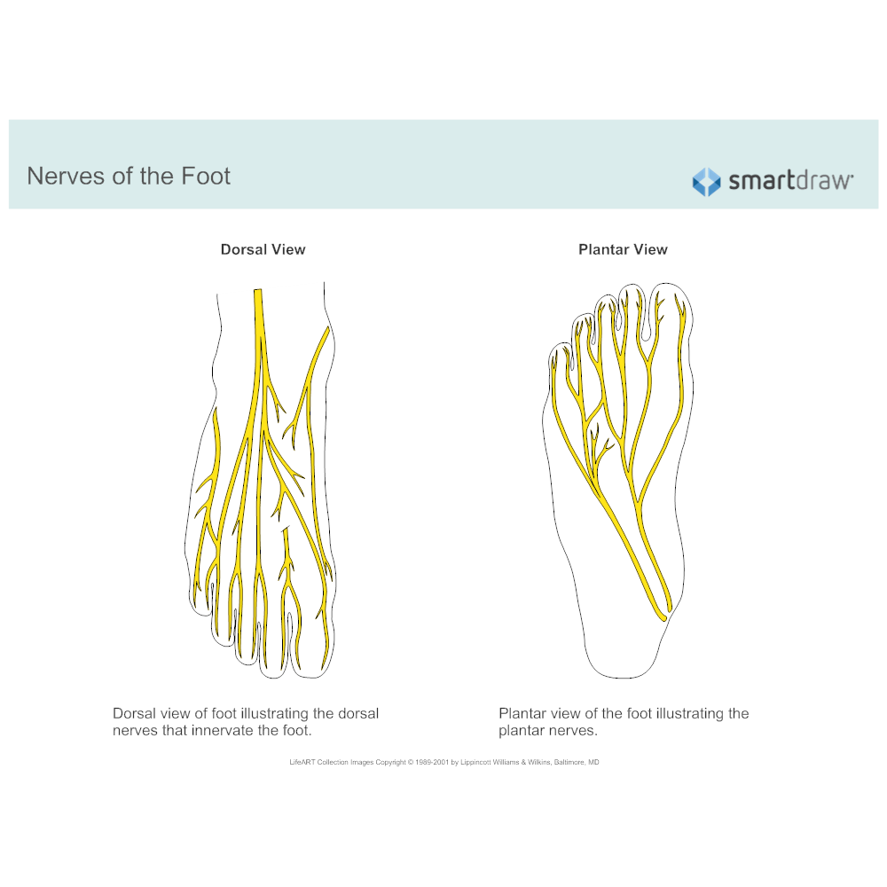
Jul 03, 2018 · All of these nerves extend as branches of nerves in the leg that pass through the ankle and into the foot. The sural nerve branches from the tibial and common fibular nerves and is responsible for feeling on the outside of the foot and the small toe. The medial and lateral plantar nerves are the two largest nerves in the bottom of the foot. Ankle and foot. The ankle region is innervated by articular branches of the tibial and deep fibular nerves. Regarding the nerves of the foot, we have the following: dorsal digital nerves, proper plantar digital nerves, lateral dorsal cutaneous nerve, and plantar (medial and lateral) nerves.
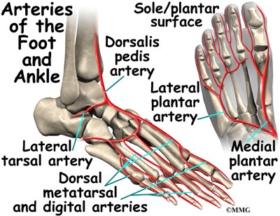
provides blood supply to plantar foot and toes. branches. plantar digital arteries. plantar metatarsal arteries. Arcuate artery. is a vascular arch that runs in the dorsal midfoot deep to the extensor tendons. gives off dorsal metatarsal arteries that run in the 2nd, 3rd and 4th intermetatarsal spaces.

Nerves of the foot and ankle diagram
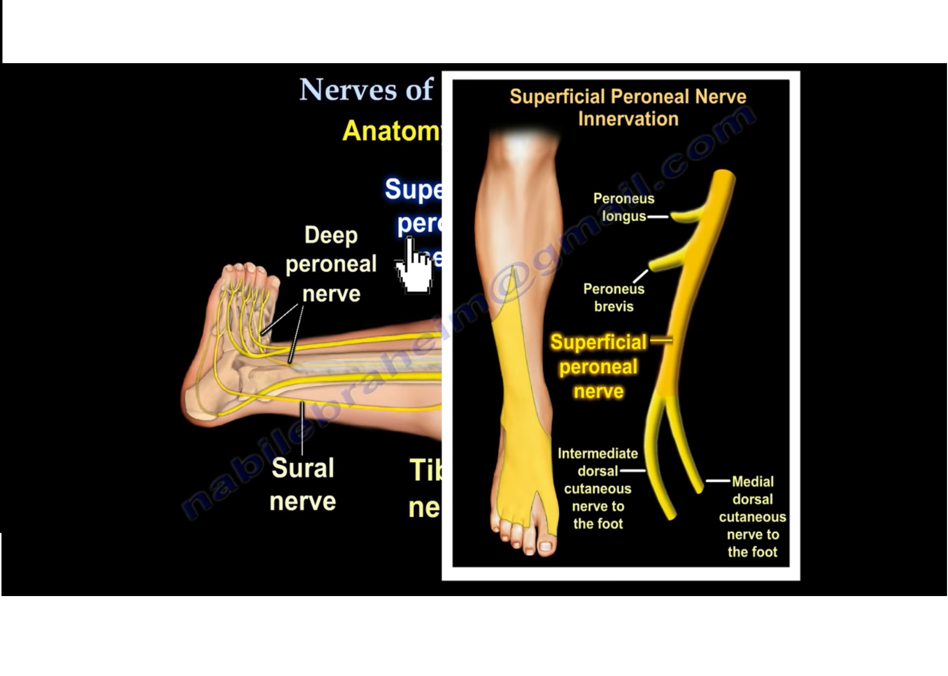
3%. (70/2732) 3. It is the terminal branch of the superficial peroneal nerve; injury leads to reduced sensation over medial aspect of great toe. 83%. (2260/2732) 4. It is the terminal branch of the deep peroneal nerve; injury leads to first interphylangeal joint flexion weakness. 3%. This nerve enters the foot along the outside of the ankle and runs to the fifth toe, supplying only the lateral side of the toe. On the opposite side of the foot is a nerve that is not counted among the dorsal digital nerves, the saphenous nerve. This nerve penetrates the medial side of the foot, ending just beneath the big toe, and it is the ... Damage to the foot and ankle tendons are a common cause of foot pain, typically caused by overuse, overstretching or an injury. Tendons are thick bands of tissue that connect muscles to bone. When a muscle contracts, the tendon pulls on the bone causing the joint to move. There are a number of tendons located in the foot and ankle all ...
Nerves of the foot and ankle diagram. The SuN, a pure sensory nerve, accesses the foot via a posterior approach to innervate the lateral aspect of the ankle through the base of the 5 th ray. This may be absent in up to 20% of individuals per cadaveric studies. 9 Tibial nerve (TN) 1,3,7 The TN is a direct continuation of the medial trunk of the sciatic nerve. partment is the posterior tibial. The tibial nerve provides the nerve supply. The ankle and foot. The 26 bones of the foot create an architectural vault, sup-ported by three arches and resting on the ground at three points, which lie at the corners of an equilateral triangle (Fig. 2). Ligaments bind the bones to provide the static stability of ... Problems with nerves in the feet are very common. Many times, an injured nerve will cause intense pain and heat felt within the foot. Nerves act as a network, communicating important information from the foot to the brain. Learn more about the various conditions and problems that can affect the nerves in the foot. Match the corresponding numbers on the foot diagram below for a list of conditions that may be causing your foot and ankle pain. This is meant for educational purposes only. If you're having a problem with your foot or ankle, visit a podiatrist - a foot and ankle specialist! Top (Dorsal) View of Foot & Ankle Number 1 and 2:
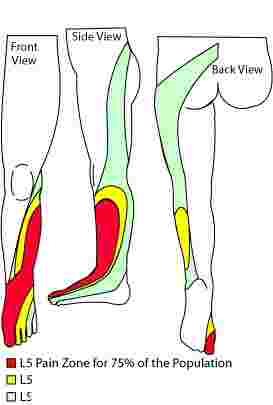
Nerves in Ankle The nerve supply of the ankle is from nerves that pass by the ankle on their way into the foot. The tibial nerve runs behind the medial malleolus. Another nerve crosses in front of the ankle on its method to top of the foot. There is also a nerve that passes along the outer edge of the ankle. A high arch is the opposite of a flat foot, and somewhat less common. The term pes cavus encompasses a broad spectrum of foot deformities. However, a pain management doctor gave me a diagram of the L5 and S1 nerve, showing they split off. He also said in spinal fusion, this is more often than not the outcome. Nerves In Foot Diagram. nerves of the leg and foot along its route through the legs the sciatic nerve splits into the tibial and mon fibular peroneal nerves which in turn split into many smaller nerves in the legs and feet the nerves of the foot help move the body and keep balance both while it's moving and at rest a plete guide to the nerves in your feet foot vitals tingling feet the ... Upon arising, the sural nerve descends between the heads of the gastrocnemius muscle. It then passes near the lateral margin of the calcaneal tendon, ( running alongside the small saphenous vein until it reaches the ankle. The sural nerve then passes between the lateral malleolus and the calcaneus and enters into the foot.
The fourth nerve of the foot is another branch of the tibial nerve, known as the sural nerve (Figure 17). This nerve runs from slightly below the knee to the lateral aspect of the foot. It becomes a very superficial nerve at the level of the posterolateral ankle and continues distally to provide sensation to the outside of the foot. It has no motor function. Download scientific diagram | Anatomical dissection of the cutaneous nerves of the foot and ankle. 1 Superficial peroneal nerve, 2 Fascial piercing of the superficial peroneal nerve, 3 Superficial ... Dr. Ebraheim's educational animated video describes the nerves of the lower leg in a very easy and simple animation.Lateral cutaneous nerve of the calfSural ... Foot Pain Diagram. Written By: Chloe Wilson BSc(Hons) Physiotherapy Reviewed By: FPE Medical Review Board A foot pain diagram is a great tool to help you work out what is causing your ankle and foot pain. There are a whole range of structures e.g. bones, muscles, tendons and nerves which will each give slightly different foot pain symptoms.
Nerve Diagram Foot - Diagram Media
The anatomy of the nerves of the foot and ankle is complex, and familiarity with the normal anatomy and course of these nerves as well as common anatomic variants is essential for correct identification at imaging. Ultrasonography (US) and magnetic resonance (MR) imaging allow visualization of these nerves and may facilitate diagnosis of various compression syndromes, such as "jogger's ...
Vessels and nerves of sole of foot
The arteriessupplying the ankle joint are branches of the anterior and posterior tibial arteries as well as the peroneal artery. The nervesof the ankle are derived from the deep and superficial peroneal nerves, the tibial nerves, and the sural and saphenous nerves. The Foot Bones of the foot as seen from the medial (arch) side.

Acute and Chronic Sciatica - Causes and removing the pain ...
The nerves on the front and outer edge of the ankle control the muscles in this area, and they give sensation to the top and outside edge of the foot. Blood Vessels. The ankle gets blood from nearby arteries that pass by the ankle on their way to the foot. The dorsalis pedis runs in front of the ankle to the top of the foot. (You can feel your ...

Femoral Impingement and Runner's Dystonia : 2019
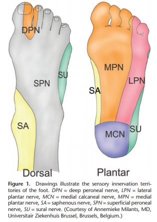
There are six nerves associated with the motor and sensory functions of the foot and ankle. They are: Nerve Innervations: Superficial peroneal nerve (L4-S1) Lateral plantar nerve (S2-S3) Medial plantar nerve (L5-S3)

Areas of the foot and their associated nerves: (1 ...
various nerves about the ankle and foot. It is also important to recognize common anatomic vari-ants. In this article, we review the normal anat-TEACHING POINTS During ankle dorsiflexion, excessive traction may occur along the exit point of the superficial peroneal nerve, injuring it and causing secondary neuroma formation at this level. The nerve

Anatomy of the Left Ankle | Trial Exhibits, Inc.
The innervation of the lower leg, ankle and foot begins upstream at the lombo-sacral region of the spine with a bundle of nerves that together form the lumbo-sacral plexus. The nerves exiting the 4th and 5th lumbar vertebrae form the lumbo-sacral trunk merge with the sacral plexus, which consists of the sacral nerves from the 1st to the 4th ...
The Deep Fibular Nerve - Course - Motor - Sensory ...
Nerves in the feet send messages, such as indications of heat and pain and other information, to the brain. The dorsal digital nerves of the foot branch throughout the body of the foot and down ...

Pin on Feet Care
The femoral nerve contributes one nerve to the ankle: 5) The saphenous nerve (L3-L4) is just anterior to the medial malleolus. Sensory: anteromedial side of the leg, medial side of foot. Technique: see below, blocked with deep peroneal n. and superficial peroneal n. The deep peroneal, superficial, peroneal, and saphenous nerves can be blocked ...

The nerves of the foot - Stock Image - F001/8178 - Science ...
Neuritis and Neuromas of the Foot and Ankle. Edited by Eric Malicky MD . Clinical Presentation. Injury or irritation to nerves in the foot and ankle often creates pain and/or numbness. They occur as a result of an injury to a specific nerve. Nerve damage can result from the original injury, casting/wrap, or surgery.
:format(jpeg)/images/anatomy_term/n-cutaneus-dorsalis-lateralis/hTNu9Za2zSmfvzSGCPFg_Nervus_cutaneus_dorsalis_lateralis_pedis_01.png)
Diagram / Pictures: Arteries and nerves of the foot ...
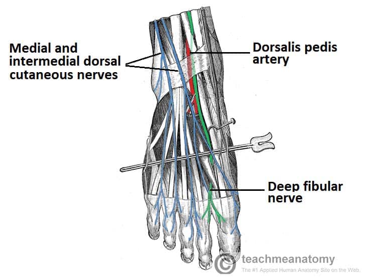
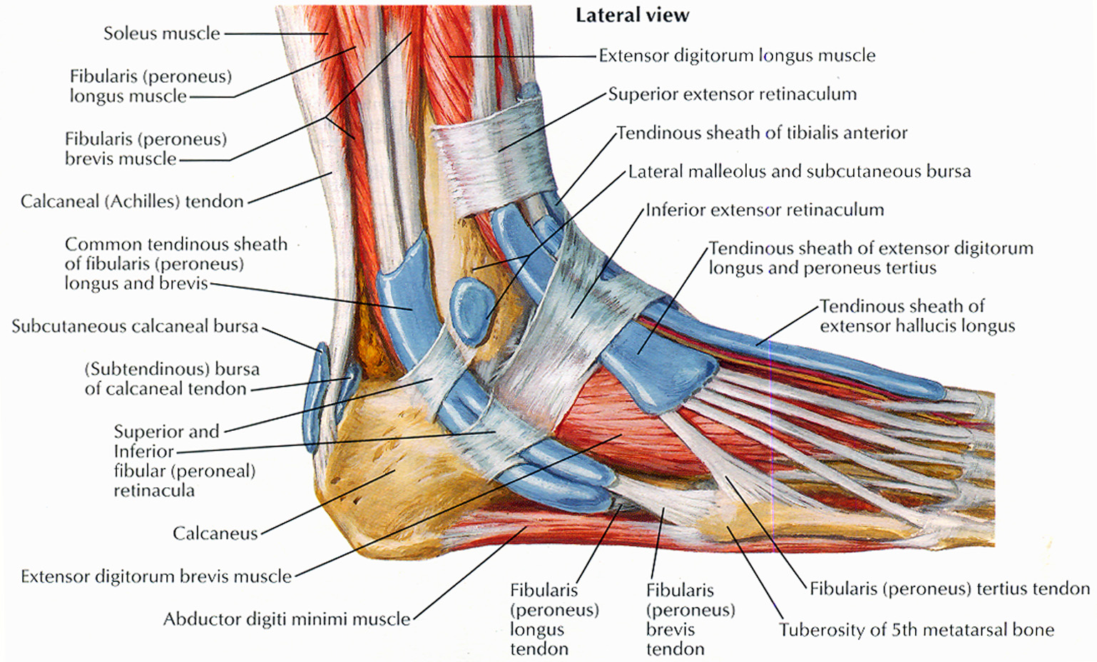
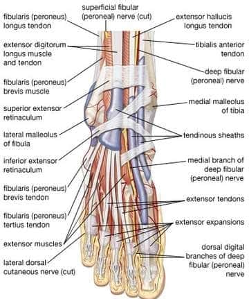
The deep fibular nerve is parallel and lateral to the tendon of the extensor hallucis longus muscle and goes inside the dorsal aspect of the foot on the lateral aspect of the dorsalis pedis artery. The nerve produces a lateral branch just distal towards the ankle joint, which stimulates the extensor digitorum brevis from its deep surface.
Ankle Anatomy | eOrthopod.com
Four common nerve problems can cause foot pain: Morton's neuroma, tarsal tunnel syndrome, diabetic peripheral neuropathy, and a pinched nerve. You'll probably know when trouble strikes. Nerve problems often trigger burning or shooting pain. And the sensation can be so intense that it can rouse you from a deep sleep.

The Tibial Nerve - Course - Motor - Sensory - TeachMeAnatomy
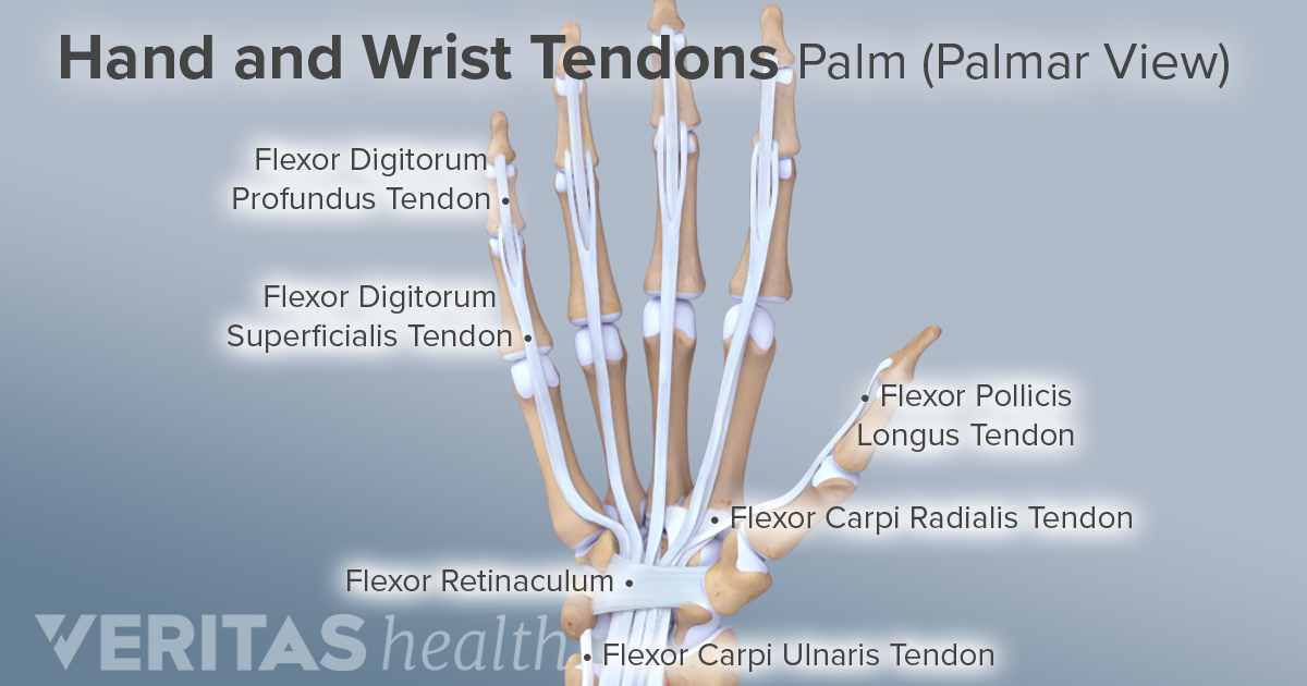
Damage to the foot and ankle tendons are a common cause of foot pain, typically caused by overuse, overstretching or an injury. Tendons are thick bands of tissue that connect muscles to bone. When a muscle contracts, the tendon pulls on the bone causing the joint to move. There are a number of tendons located in the foot and ankle all ...
Foot pain nerve, how to get rid of corns on toes in a week
This nerve enters the foot along the outside of the ankle and runs to the fifth toe, supplying only the lateral side of the toe. On the opposite side of the foot is a nerve that is not counted among the dorsal digital nerves, the saphenous nerve. This nerve penetrates the medial side of the foot, ending just beneath the big toe, and it is the ...
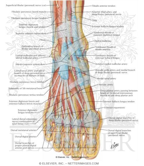
Muscles, Arteries, and Nerves of Front of Ankle and Dorsum ...
3%. (70/2732) 3. It is the terminal branch of the superficial peroneal nerve; injury leads to reduced sensation over medial aspect of great toe. 83%. (2260/2732) 4. It is the terminal branch of the deep peroneal nerve; injury leads to first interphylangeal joint flexion weakness. 3%.

Nerves of the Foot and Ankle

Frog, Deep Sea World, North Queensferry, Fife, Scotland.
Nerves Of Foot Unlabeled. Stock Photo 228843271 : Shutterstock
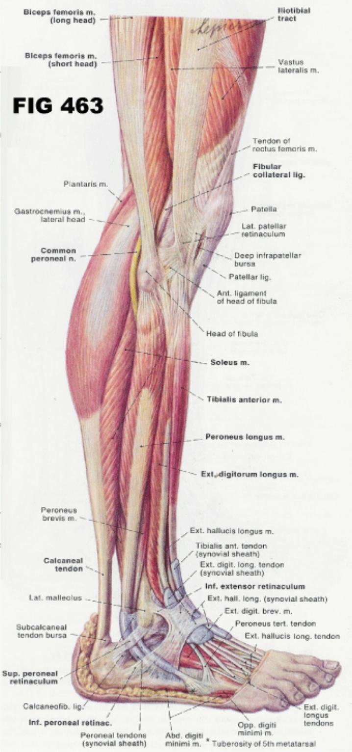
Muscular Anatomy
Nerves of the Leg and Foot — OrthopaedicPrinciples.com
Muscles that lift the Arches of the Feet

113 Best Foot anatomy images | Drawing tips, Feet drawing ...

monochrome persons feet

Injuries That Commonly Occur with a Stinger
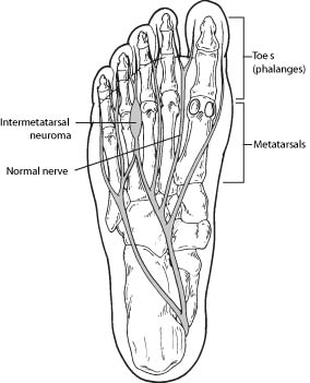
Morton's Neuroma (Intermetatarsal Neuroma) - Foot Health Facts
Ankle block : Nerves and Landmarks | Epomedicine

5 Dissection of the medial ankle, from Bodies, The ...
Ligaments, Tendons, and Nerves of the Wrist

Preoperative picture showing foot drop following common ...

Nerves of the foot, illustration - Stock Image - F029/7003 ...

Take a super ball, put it on the floor and rub your foot ...

Ligaments Of The Foot | Muscles, Tendons & Ligaments of ...

Nerves of the plantar foot | MyFootShop.com

Human Anatomy Scientific Illustrations Foot Nerves High ...

Diagram Showing Parts Of The Foot
Nerves in Foot and Ankle | IYTmed.com

Image from page 88 of "An illustrated encyclopædic medical dictionary. Being a dictionary of the technical terms used by writers on medicine and the collateral sciences, in the Latin, English, French and German languages" (1890)
Nerves of the Foot
Mixed cranial nerves: Anatomy, course, fibers, functions ...

Warm bath before treatment
Tingling in feet and legs faces the chiropractor on a ...

Nerves of the Foot and Ankle

Barefoot at the beach
0 Response to "42 nerves of the foot and ankle diagram"
Post a Comment