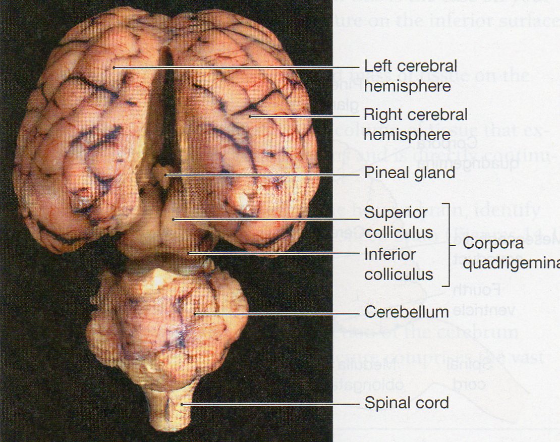
41 sheep brain diagram labeled
Sheep Brain Dissection Guide - Contents - StuDocu Sheep brains are used in this lab because they are easy to extract, reasonably inexpensive (they are procured from the food industry), large, and mammalian. A structure is anterior to another structure when it is closer to the nose of an animal (see the above diagram). Some texts use the terms anterior... PDF Sheep Neuroanatomy Lab- Labeling Worksheet Sheep Neuroanatomy Lab- Labeling Worksheet. Psychology 2315- Brain and Behaviour Kwantlen Polytechnic University. Figure 1: Dorsal view.
18 Inspirational Sheep Kidney Dissection Labeled Sheep Kidney Dissection Labeled to view on Bing2 20Feb 21 2013 This is a guide to help students at Northern Arizona University to dissect a sheep kidney mooreschools cms lib OK01000367 Centricity Web viewKidney Diagram Drawing Check Off List Renal Sinus Hilus Ureter Renal Artery Renal Vein...

Sheep brain diagram labeled
PDF Page 1 text continues here | Teacher Guide Sheep Brain Dissection Lesson Summary: Dissecting a sheep brain, students gain. appreciation for the complexity of the brain. Label the major parts and explain their functions on a picture of a sheep and/or human brain. • Sheep Brain Dissection Project Guide | HST Learning Center The sheep brain is exposed and each of the structures are labeled and described in a sequential manner, in the same way that a real dissection Learn the external and internal anatomy of sheep brains with HST's Learning Center science lesson and guide! Diagram worksheets also included. learning-center.homesciencetools.com › articleSheep Heart Dissection Lab for High School Science | HST By studying the sheep’s anatomy, you can learn how your own heart pumps blood through your body, thereby keeping you alive! Use this sheep heart dissection guide in a lab for high school students. You can also look at the labeled pictures to get an idea of what the heart looks like (that’s especially helpful for younger students).
Sheep brain diagram labeled. labeled diagram of brain- midsagittal view | Diigo Groups Sagital Cut Sheep Brain Labeled | sheep brain map midsaggital view labeled diagram of brain midsagittal. In this lab guide, students are given instruction on how to remove the dura mater, and locate the main structures of the external and internal brain. Sheep Brain Dissection with Labeled Images The sheep brain is exposed and each of the structures are labeled and described in a sequential manner, in the same way that a real dissection would occur. Listing Of Websites About sheep brain diagram labeled. The Sheep Brain Atlas at Michigan State University The Sheep Brain Atlas. John I. Johnson, Keith D. Sudheimer, Kristina K. Davis, Garrett M. Kerndt, and Brian M. Winn Radiology Department, Neuroscience Program,and Communications Technology Laboratory, Michigan State University, East Click on the labels to view the glossary definitions. Diagram of Sheep Brain Dissection labeled | Quizlet Start studying Sheep Brain Dissection labeled. Learn vocabulary, terms and more with flashcards, games and other study tools. Only RUB 193.34/month. Sheep Brain Dissection labeled.
Sheep Brain Dissection - DocsBay Sheep brain dissection: pre-lab. (Pre-Lab must be submitted to start the lab). Part 1: Planes and Axis of the Brain. 1. Label the diagram to the right with the dorsal-ventral axis and the anterior-posterior axis. 2. Name the view of the brain shown in the diagrams below en.wikipedia.org › wiki › HypothalamusHypothalamus - Wikipedia Fos-labeled cell analysis showed that the PMDvl is the most activated structure in the hypothalamus, and inactivation with muscimol prior to exposure to the context abolishes the defensive behavior. Therefore, the hypothalamus, mainly the PMDvl, has an important role in expression of innate and conditioned defensive behaviors to a predator. Labeled Brain Model Diagram | Science Trends The brain is so complex that even to simply discuss it, certain distinctions have to be made regarding its structure, and for that reason, scientists divide the brain up into three major portions with each of these portions divided into many subregions and structures. en.wikipedia.org › wiki › ClitorisClitoris - Wikipedia The clitoris (/ ˈ k l ɪ t ər ɪ s / or / k l ɪ ˈ t ɔːr ɪ s / ()) is a female sex organ present in mammals, ostriches and a limited number of other animals.In humans, the visible portion – the glans – is at the front junction of the labia minora (inner lips), above the opening of the urethra.
Sheep Brain Dissection Project Guide | HST Learning Center Diagram worksheets also included. Sheep Brain Dissection Guide Project. Sheep brains, like other sheep organs, are much smaller than human brains but have similar features. Use this for a high school lab, or just look at the labeled images to get an idea of what the brain looks like. sheep brain labeled brain lateral view04 - Made By Creative Label {Label Gallery} Get some ideas to make labels for bottles, jars, packages, products, boxes or classroom activities for free. An easy and convenient way to make label is to generate some ideas first. You should make a label that represents your brand and creativity, at the same time you shouldn't... sheep brain lab dissection Then label the internal anatomy diagram with all the structures. (Note: the pituitary gland is usually not visible on the sheep brain) corpus callosum, medulla oblongata, pituitary gland, ventricle, pons, sulci, gyri, cerebral. cortex, thalamus, hypothalamus, cerebellum, pineal gland. DOC Sheep Brain Anatomy Lab Manual The brain of the sheep is useful for study because its anatomy is similar to human brain anatomy. Although exact proportions (and names) sometimes differ The cruciate fissure (labeled ansate sulcus in your photo atlas) is known in the human brain as the fissure of Rolando or central sulcus, and...
35 Blank Brain Diagram To Label - Label Design Ideas 2020 The nervous system an image of the nerves of the lower body with blank labels attached. 8 in the diagram 1. Label Br...
Diagram of Sheep Brain - Lateral view
› parasitology › hostHost-Parasite Relationship (With Diagram) - Biology Discussion ADVERTISEMENTS: Parasitism is an association or a situation in which two organisms of different taxonomic positions live together where one enjoys all sorts of benefits (like derivation of nourishment, reproduction etc. which are basic requirements for existence) at the expense of the other. The benefited organism is called the parasite and the organism harbouring the […]
PDF Sheep Brain Explora/on Guide Sheep Brain Explora/on Guide. Why dread a bump on the head? Lesson 2: What does the brain While we are using the sheep brain for dissection, this guide will highlight the similarities between the Label the following anatomical directions in the previous figure: rostral, caudal, dorsal, lateral...
PDF Label the Parts of a Sheep Brain Sheep Brain Dissection. Sheep brains, although much smaller than human brains, have similar Use this as a dissection guide complete enough for a high school lab, or just look at the labeled images to get an Print out these diagrams and fill in the labels to test your knowledge of sheep brain anatomy.
Sheep Brain Diagram Labeled - Free Catalogs A to Z 6 hours ago Image Result For Sheep Brain Labeled Brain Diagram Human Brain Diagram Brain Anatomy. Sheep Brain Dissection Project Guide Hst Learning Center Dissection Brain Mapping Science Biology.
Sheep Brain Dissection Bi - BIOLOGY JUNCTION Sheep Brain Dissection. Introduction: Objective: Materials: Procedure: Label The Following: 7. cerebellar vermis 8. cerebellar hemisphere 9. pyriform lobe 10. rhinal fissure 11. lateral olfactory tract.
Sheep Brain Dissection with Labeled Images The sheep brain is exposed and each of the structures are labeled and described in a sequential manner, in the same way that a real 1. The sheep brain is enclosed in a tough outer covering called the dura mater. You can still see some structures on the brain before you remove the dura mater.
› muhamadalhakimasri › frogFrog dissection lab answer key - SlideShare Jul 16, 2015 · Posterior to the cerebellum is the medulla oblongata (E) this is the which connects the brain to the spinal cord (F). To receive extra credit for exposing the brain you must first present a completed the data table and have all the brain parts labeled then show the brain dissection to your teacher for approval. The cleaner the dissection the ...
Sheep Brain Dissection Guide Good Luck!! Ms. Lecce 6. Main Brain Regions • Identify the cerebrum, brain stem) and cerebellum on your brain • Draw picture of sheep brain and label structures Cerebellum Cerebrum Brain stem. Label your diagram with parts of midbrain and brain stem • What is the function of the medulla oblongata and pons? •
Sheep Brain Images Additional Resources. Nervous system - sheep brain images. Sheep Brain Labeled.
Sheep Brain & Eye (with labels) - YouTube A video tutorial of the anatomy of the brain and eye of a sheep for comparative anatomy.
Lab Exam 3: Anatomy of Sheep Brain; Histology Flashcards - Cram.com Diagram of sheep brain, dorsal view. Neuron cells (400x). Nerve and blood vessels slide (closeup of nerve). more nerves and blood vessels. artery vein nerve diagram. note that some axons are myelinated and others are unmyelinated. review: what type of tissue is on the slide.
Sheep Brain - Median1 View Sheep Brain — Medial View. In this median view of a sheep brain, labeled telencephalic structures include: rostral commissure (black thread) located in the lamina terminalis; corpus callosum (extending from 1 to 2) and cingulate gyrus (4). Also labeled are: pineal body (blue pic), the interthalamic...
› jdownloads › GET_TaskGRADE: 5 SUBJECT: NATURAL SCIENCES AND TECHNOLOGY TERM ONE ... B. Sheep C. Lion D. Giraffe. (1) (2) Question 2 Match the definitions in column A with the word in column B. Write the letter from column B as your answer in the middle column. 2. COLUMN A Answer COLUMN B 2.1 Begins to sprout or grow into a seed A. Habitat 2.2 The place where two or more bones meet B. Germination
anatomylearner.com › chicken-skeleton-anatomyChicken Skeleton Anatomy with Labeled Diagram ... Aug 22, 2021 · Chicken skeleton labeled diagram. Thanks for continuing to learn chicken skeleton with a labeled diagram. Here, I tried to show you every single bone from a chicken. Unlike mammals, you will find axial and appendicular structures in a chicken or a bird.
PDF Dissection of the sheep's brain Caudal. Sheep Brain Dissection Guide. Planes of Orientation In addition to the direction, the brain as a three dimensional object can be divided. In addition you should see the crossing of the anterior commissure right above the optic chiasm. While not labeled see if you can see the corpus callosum...
Sheep brain dissection | Human Anatomy and Physiology Lab... The sheep brain is quite similar to the human brain except for proportion. The sheep has a smaller cerebrum. Examining the internal sheep brain. Use a knife or a scalpel to cut the specimen along the longitudinal fissure. This will allow you to separate the brain into the left and right hemispheres.
Image result for sheep brain labeled | Brain diagram, Human brain... Human Brain Anatomy. Image result for sheep brain labeled. Image of the brain showing its major features for students to practice labeling. Answers are included.
learning-center.homesciencetools.com › articleSheep Heart Dissection Lab for High School Science | HST By studying the sheep’s anatomy, you can learn how your own heart pumps blood through your body, thereby keeping you alive! Use this sheep heart dissection guide in a lab for high school students. You can also look at the labeled pictures to get an idea of what the heart looks like (that’s especially helpful for younger students).
Sheep Brain Dissection Project Guide | HST Learning Center The sheep brain is exposed and each of the structures are labeled and described in a sequential manner, in the same way that a real dissection Learn the external and internal anatomy of sheep brains with HST's Learning Center science lesson and guide! Diagram worksheets also included.
PDF Page 1 text continues here | Teacher Guide Sheep Brain Dissection Lesson Summary: Dissecting a sheep brain, students gain. appreciation for the complexity of the brain. Label the major parts and explain their functions on a picture of a sheep and/or human brain. •

0 Response to "41 sheep brain diagram labeled"
Post a Comment