44 atomic force microscope diagram
Atomic force microscopy is arguably the most versatile and powerful microscopy technology for studying samples at nanoscale. It is versatile because an atomic force microscope can not only image in three-dimensional topography, but it also provides various types of surface measurements to the needs of scientists and engineers.
File:Atomic force microscope block diagram.svg. Size of this PNG preview of this SVG file: 646 × 599 pixels. Other resolutions: 259 × 240 pixels | 517 × 480 pixels | 647 × 600 pixels | 828 × 768 pixels | 1,104 × 1,024 pixels | 926 × 859 pixels.
Berkeley Lab scientists make the first-ever high-resolution images of a molecule as it breaks and reforms chemical bonds. News Release Paul Preuss 510-486-6249 • May 30, 2013. Almost as clearly as a textbook diagram, this image made by a noncontact atomic force microscope reveals the positions of individual atoms and bonds, in a molecule ...

Atomic force microscope diagram
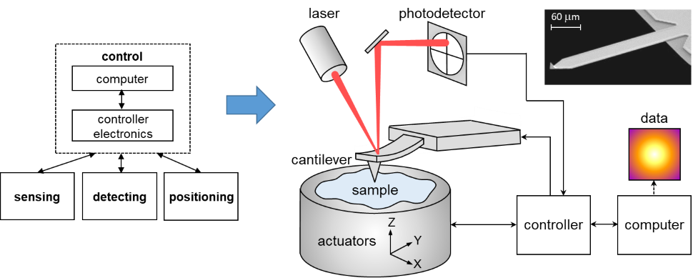
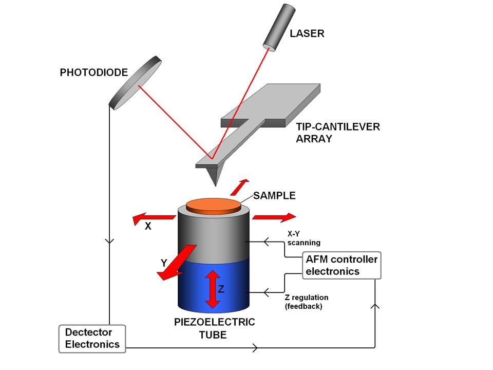
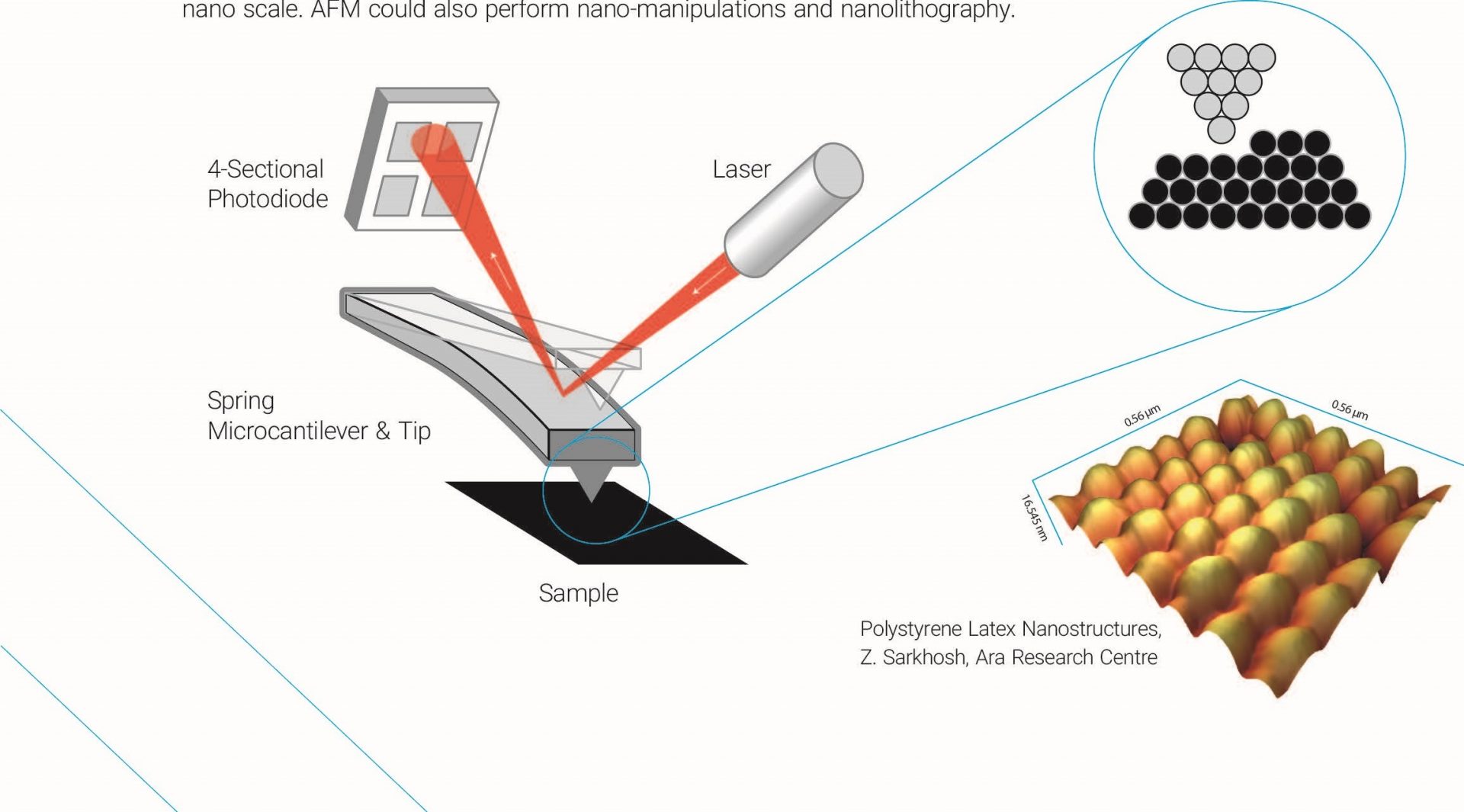
AFM, which uses a sharp tip to probe the surface features by raster scanning, can image the surface topography with extremely high magnifications, up to 1,000,000X, comparable or even better than electronic microscopes. The measurement of an AFM is made in three dimensions, the horizontal X-Y plane and the vertical Z dimension.
13.2.4.4 Atomic force microscope. AFM is a kind of scanning probe microscope which is used to calculate properties such as the height, magnetic force, surface potential, and friction, and also has the ability to measure intermolecular forces.
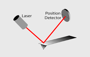
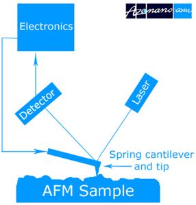
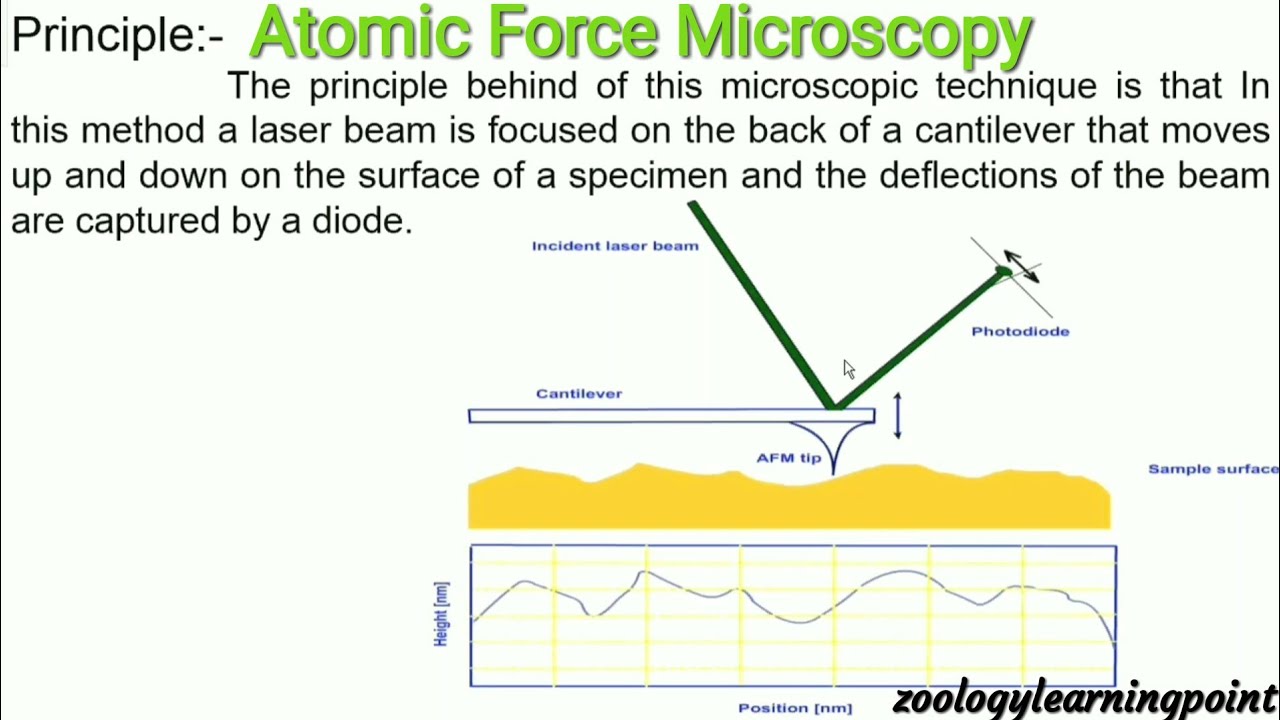
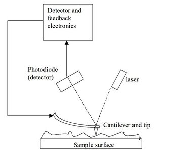
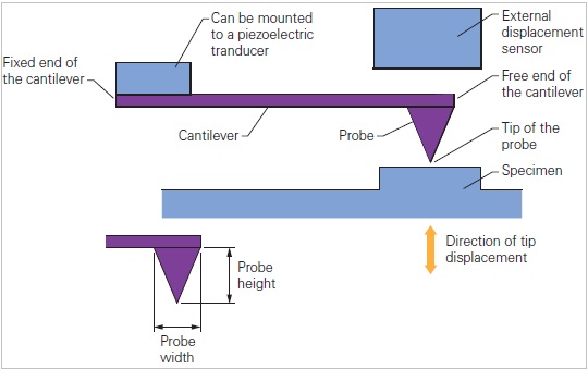
In this article we will discuss about the design of atomic force microscope, explained with the help of a diagram. An atomic force microscope instead of using a lens is provided with a probe to examine the surface of a specimen with a sharp tip which may be several micrometers in and less than 10 nm in diameter at the point near field to be examined. The tip lies at the end of the lever which ...
Atomic force microscope diagram.
Functionalizing the tip of an atomic force microscope with a CO molecule enabled atomic-resolution imaging of single molecules, and measurement of their adsorption geometry and bond-order relations. In addition, by using scanning tunneling microscopy and Kelvin probe force microscopy, the density of the molecular frontier orbitals and the ...
Atomic Force Microscopy (AFM) 1. General Principle The Atomic Force Microscope is a kind of scanning probe microscope in which a topographical image of the sample surface can be achieved based on the interactions between a tip and a sample surface. The atomic force microscope was invented by Gerd Binning et al. in 1986 at IBM Zurich based on ...
Scanning probe microscopy has been the engine of characterization in nanoscale systems ().Atomic force microscopy (AFM) in particular has developed into a leading technique for high-resolution studies without material restrictions (3-5).It is increasingly being used for detailed characterization in a wide variety of physical, biological, and chemical processes (6, 7).
Keywords:Atomic force microscopy, Protein structure, Amyloid, Disease, Correlative microscopy, NMR. Abstract:Proteins are versatile macromolecules that perform a variety of functions and participate in virtually all cellular processes. The functionality of a protein greatly depends on its structure and alterations may result in the development ...
§D. Sarid, Scanning Force Microscopy with Applications to Electric, Magnetic and Atomic Forces , Revised Edition, Oxford University Press, 1994. § U. Dürig, "Interaction sensing in dynamic force microscopy", New Journal of
a Single-Molecule Level by Atomic Force Microscopy 179 A B Fig. 2. AFM force spectroscopy: (A) Diagram of a single stretching experiment; (B) Ex ample of a force extension curve, with definitions of force and extension. (Adapted and modified from reference 4, with permission) off the back of the cantilever onto the center of the PSPD.
Atom Under The Microscope Electron & Atomic Force Microscopy What is an Atom? Essentially, an atom is the smallest unit of an element that retains the properties of the same element (iron, copper, carbon etc). This means that divided further, its components (electrons, protons, and neutrons) do not retain the properties of the element.
Atomic Force Microscopy [AFM] Cathodoluminescence Image Analysis of Particles Micro-X-ray Fluorescence Raman Microscopy Physisorption Surface Area Spectroscopy Cathodoluminescence Spectroscopy Cathodoluminescence Spectroscopy Electron Microscope SEM, ESEM, SEM-FIB, (S)TEM EM Add-on detector SEM-Cathodoluminescence (SEM-CL) …
To learn more about Atomic Force Microscopy, click through. AFM microscopes are among the best solutions for measuring the nanoscale surface metrology and material properties of samples. A conventional compound light microscope is limited to a maximum sample magnification of approximately 1000x; a quantity that is dictated by the wavelengths of ...
The atomic force microscope's ability to measure conductive or non-conductive samples in air allows for characterization of complex polymers and biological samples. For samples that need to be kept hydrated or in a controlled liquid or pH solution, AFMWorkshop offers a fluid cell option that allows for AFM analysis in liquid.
25.06.2015 · An atomic force microscopy (AFM) image of the as-synthesized MoS 2 sheet on a Si substrate shown in Fig. 1a indicates a smooth surface topography, combined cross-sectional and image histogram ...
Download scientific diagram | Schematic drawing of the atomic force microscope. from publication: Direct Measurement of Interaction Forces between Surfaces in Liquids Using Atomic Force Microscopy ...
Sampling protein form and function with the atomic force ...
Roughness affects various part characteristics, including the amount of wear, the ability to form a seal when the part makes contact with something, and the ability to coat the part. KEYENCE's Introduction to "Roughness" website introduces parameters and case studies related to such surface measurements.
Atomic force microscopy - nanoscience instruments
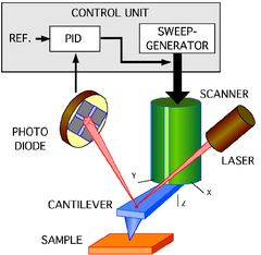
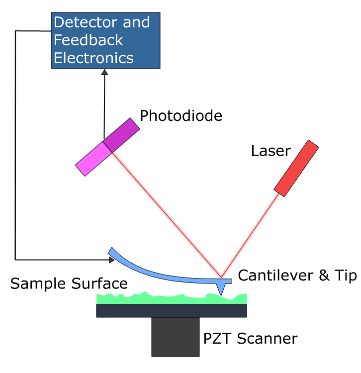
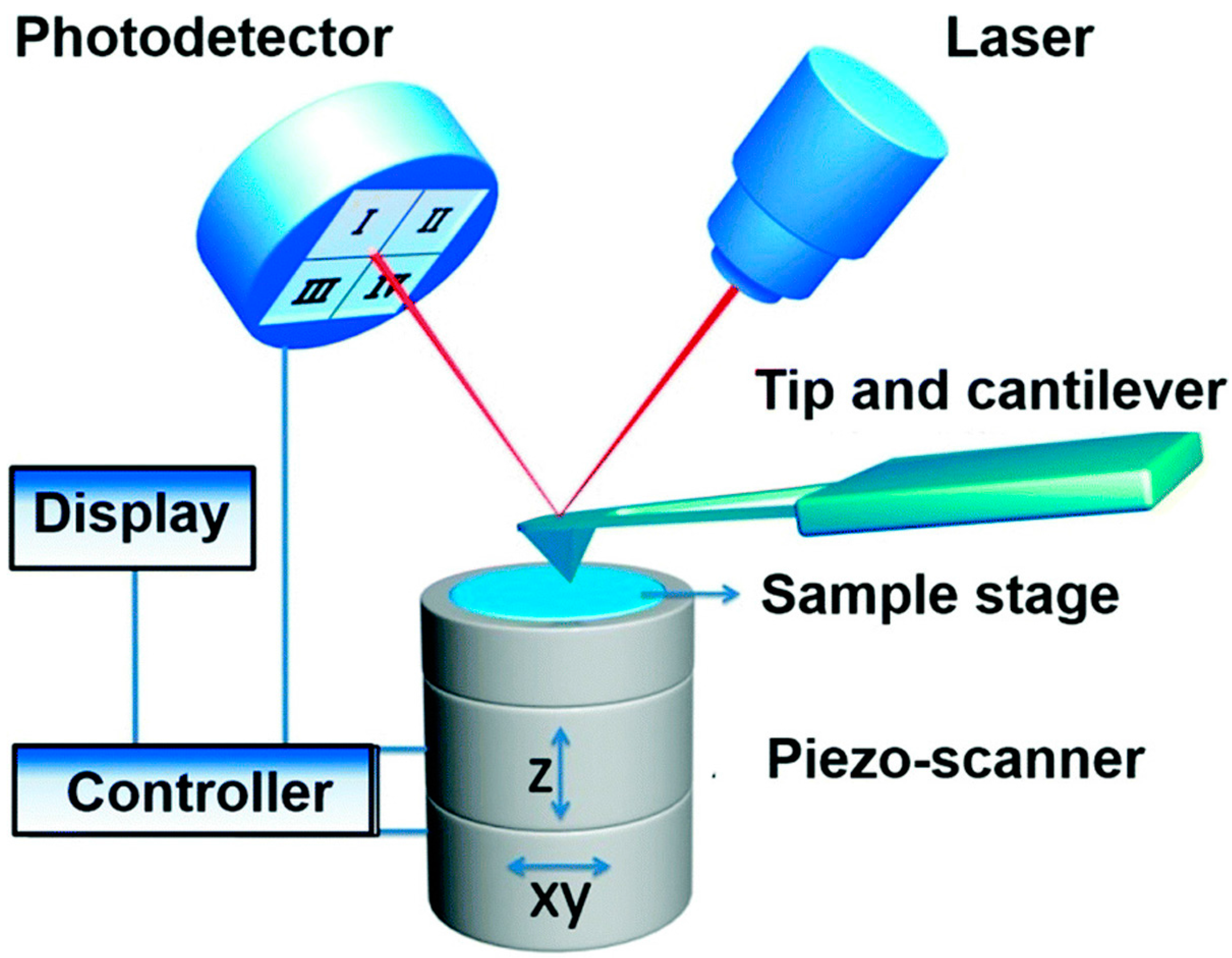
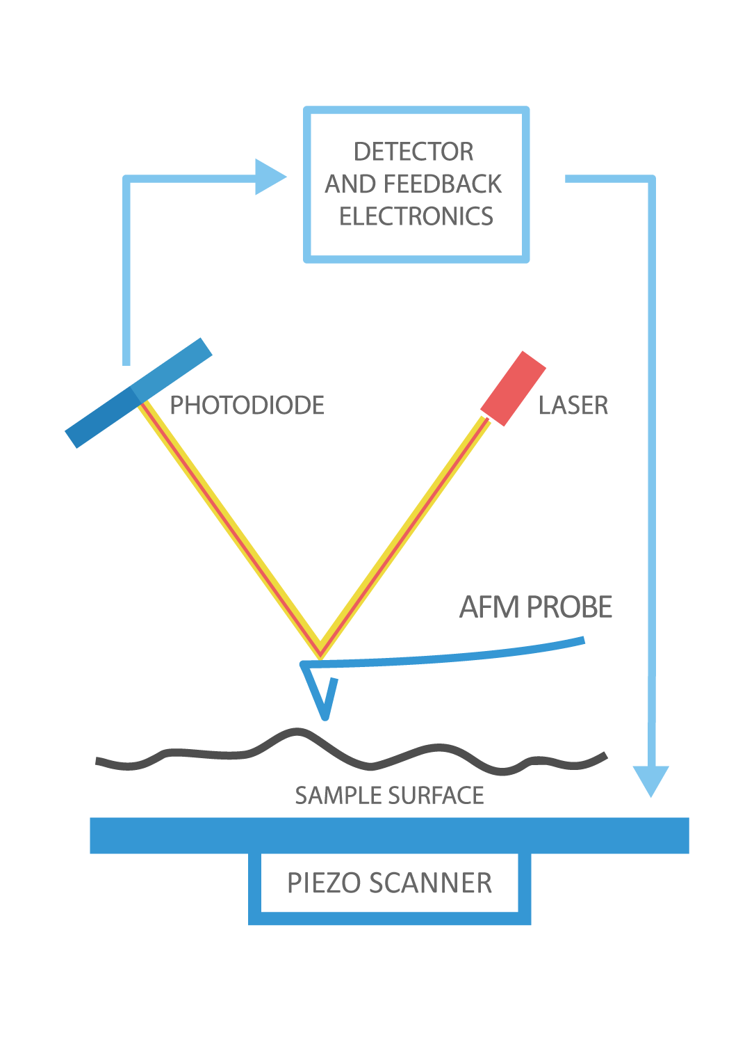
Similarly in atomic force microscopes, depending on the different modes, there is a parameter that serves as the setpoint. For example, in static mode (contact mode) the feedback parameter is cantilever deflection, while in the most common form of tapping mode, the cantilever oscillation amplitude is the feedback parameter. The instrument is ...

Mikroskop gaya atom, mikroskop optik, konduktif atomic force ...
Atomic force microscopy (AFM) is a powerful technique that enables the imaging of almost any type of surface, including polymers, ceramics, composites, glass and biological samples. AFM is used to measure and localize many different forces, including adhesion strength, magnetic forces and mechanical properties.

Upt laboratorium terpadu will be equipped with facility of ...
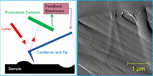
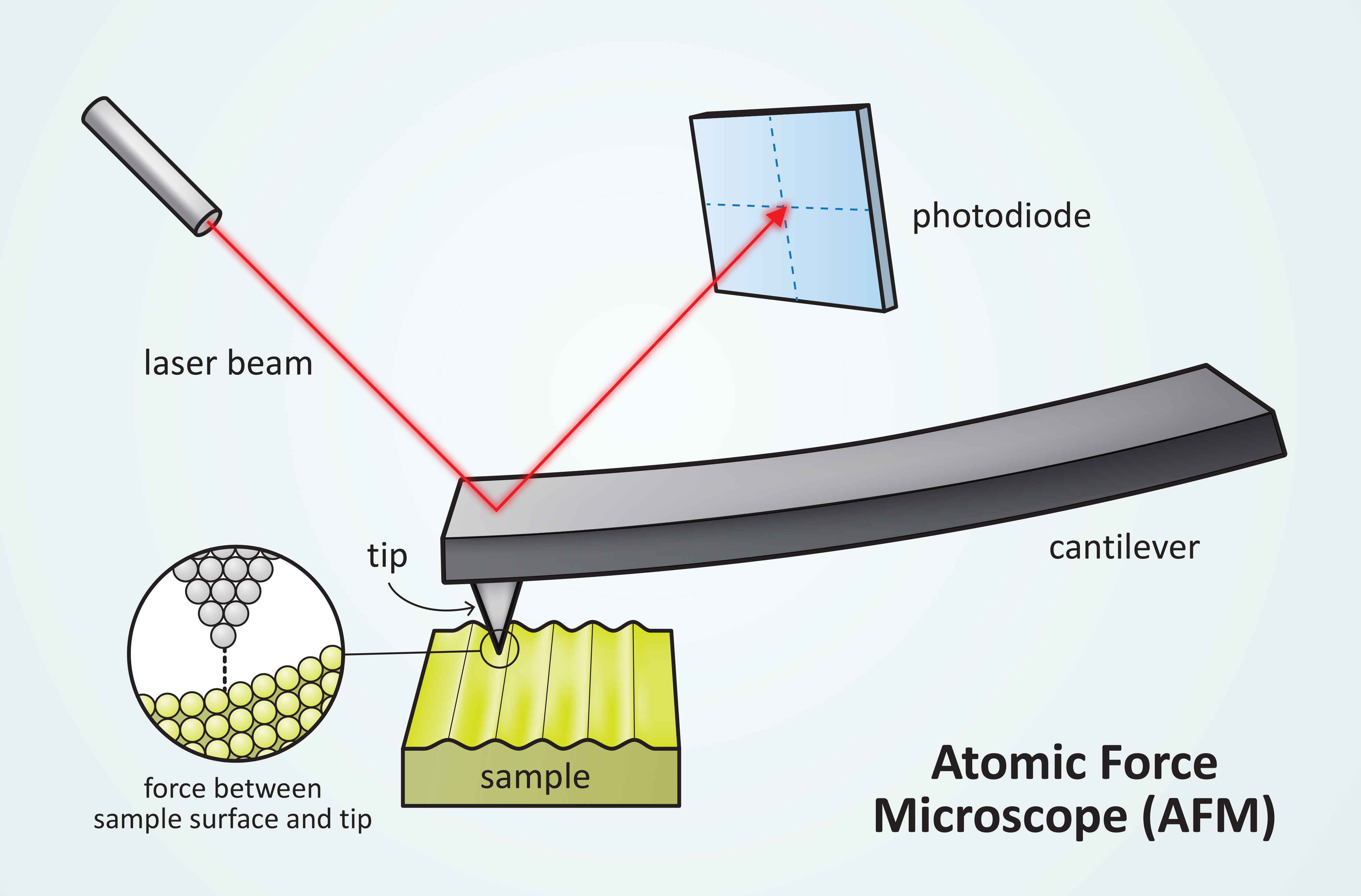
In manipulation, the forces between tip and sample can also be used to change the properties of the sample in a controlled way, for example, atomic manipulation, scanning probe lithography and local stimulation of cells. Figure 1. Block diagram of atomic force microscope using beam deflection detection.

Types of scanning probe microscopy – advanced materials 2019 ...
Whereas most atomic-level imaging is done with a scanning tunneling microscope, these images were captured with noncontact atomic force microscope (nc-AFM, pictured below).
Bone biology and mechanics lab: bbml
Focused ion beam, also known as FIB, is a technique used particularly in the semiconductor industry, materials science and increasingly in the biological field for site-specific analysis, deposition, and ablation of materials.A FIB setup is a scientific instrument that resembles a scanning electron microscope (SEM). However, while the SEM uses a focused beam of …

Karakterisasi atomic force microscope (afm) | kata pengetahuan
Atomic-force microscopy is a reference method for traceable and correlative measurements of nanostructures. The Nanostructure Fabrication and Measurement Group is developing critical-dimension and traceable microscope systems to calibrate probe tips and microscopy standards, and measure diverse devices ranging from waveguides to nanoparticles.
Atomic force microscope - fbswiki
Structure. 17(8): 1137-47. Rosenzweig R, Osmulski PA, Gaczynska M, and Glickman MH: (2008) The central unit within the 19S regulatory particle of the proteasome. Nat Struct Mol Biol. 15(6): 573-80. Gaczynska M and Osmulski PA: (2008) Chapter 3 Atomic force microscopy as a tool to study the proteasome assemblies
Sensors | free full-text | review: cantilever-based sensors ...
Atomic force microscopy (AFM) is a microscopy technique that uses a sharp probe to trace a sample surface at nanometre resolution. For biological applications, one of its key advantages is its ability to visualize substructure of single molecules and molecular complexes in an aqueous environment.
Atomic force microscopes (afm) what they are, how they work ...
The observation of "subatomic features" by atomic force microscopy (AFM) in experiments where the front atom of the tip was imaged by the highly localized dangling bonds of the Si(111)-(7x7) surface atoms raised discussions within the community ().Similar observations were reported later for Si adatoms imaging Si tips (), CoSm tips (), and even in scanning tunneling microscopy (STM) ().
File:atomic force microscope block diagram.png - wikimedia ...
Atomic force microscopy (AFM) is a type of scanning probe microscopy (SPM), with demonstrated resolution on the order of fractions of a nanometer, more than 1000 times better than the optical diffraction limit.The information is gathered by "feeling" or "touching" the surface with a mechanical probe. Piezoelectric elements that facilitate tiny but accurate and precise movements on (electronic ...
Afm/atomic force microscopy
This is a retouched picture, which means that it has been digitally altered from its original version.Modifications: translation to german.The original can be viewed here: Atomic force microscope block diagram.svg: .Modifications made by Cepheiden.
Atomic force microscopy (afm) | springerlink
02.11.2018 · N Goalby chemrevise.org 1 Ionic bonding is the strong electrostatic force of attraction between oppositely charged ions formed by electron transfer. Metal atoms lose electrons to form +ve ions. Non-metal atoms gain electrons to form -ve ions. Mg goes from 1s2 2s2 2p63s2 to Mg2+ 1s2 2s2 2p6 O goes from 1s2 2s2 2p4 to O2-1s2 2s2 2p6 Ionic bonding …
Atomic force microscope
A high-speed atomic force microscope (HS-AFM) requires a specialized set of hardware and software and therefore improving video-rate HS-AFMs for general applications is an ongoing process.

Atomic force microscope (afm), the key tool for surface ...
29.10.2021 · Microscope, Microscope Parts, Labeled Diagram, and Functions Published by Admin on June 1, 2021 June 1, 2021. What is Microscope? Microscope is derived from Ancient Greek words and composed of mikrós, “small” and skopeîn,”to look” or “see”. It is one of the most revolutionized scientific instruments used to observe or examine minute structures not clearly …
Atomic force microscopy - an overview from asylum research
An Atomic Force Microscope (AFM) captures the image of the topography of the specimen by force, by scanning the cantilever across the area of significance. Based on the degree of elevation or deep the topography of the specimen is it will determine how the beam is deflected which is tracked by the positive-sensitive photo-diode (PSDP).

A schematic diagram of an atomic force microscope. | download ...
08.10.2007 · What are the parts of an atom? Most atoms have three different subatomic particles inside them: protons, neutrons, and electrons.The protons and neutrons are packed together into the center of the atom (which is called the nucleus) and the electrons, which are very much smaller, whizz around the outside.When people draw pictures of atoms, they show the …

Explain construction and working of atomic force microscope ...
Parts of Atomic Force Microscope. Atomic Force Microscopes have several techniques for measuring force interactions such as van der Waals, thermal, electrical and magnetic force interactions for these interactions done by the AFM, it has the following parts that assist in controlling its functions.
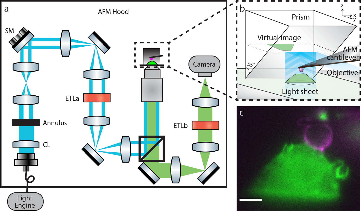
Combined atomic force microscope and volumetric light sheet ...

The atomic force microscope – probing to “see” at the ...

Block diagram of atomic force microscopy operation ...
Atomic force microscope (afm) - department of pharmacology

Atomic force microscopy - wikipedia
Scientific image - atomic force microscope illustration ...

Atomic force microscopy-based force measurements on animal ...

Schematic drawing of the atomic force microscope. | download ...

Atomic force microscopy - lnf wiki

Share ur notes: atomic force microscopy(afm) ppt
.jpg)
The principles of operation of an atomic force microscope (afm)
Sensors | free full-text | progress in the correlative atomic ...

Polymer characterization with the atomic force microscope ...

Explain the construction and working of of atomic force ...
Atomic force microscope (afm)- definition, principle, parts ...
Contact mode and tappingmode atomic force microscopy
What is afm? learn about atomic force microscopy! - nanoandmore

Laporan wawasan pasar global atomic force microscopy (afm ...
Schematic of an atomic force microscope | nist

Afm scanning modes – max iv

Schematic of an atomic force microscope. | download ...

Schematic representation of an atomic force microscope (afm ...

File:schematic of an atomic force microscope-ta.svg ...

9.2: atomic force microscopy (afm) - chemistry libretexts
Atomic force microscope ( spm complete system) - labtechniche
0 Response to "44 atomic force microscope diagram"
Post a Comment