42 in the diagram, where is the osteon?
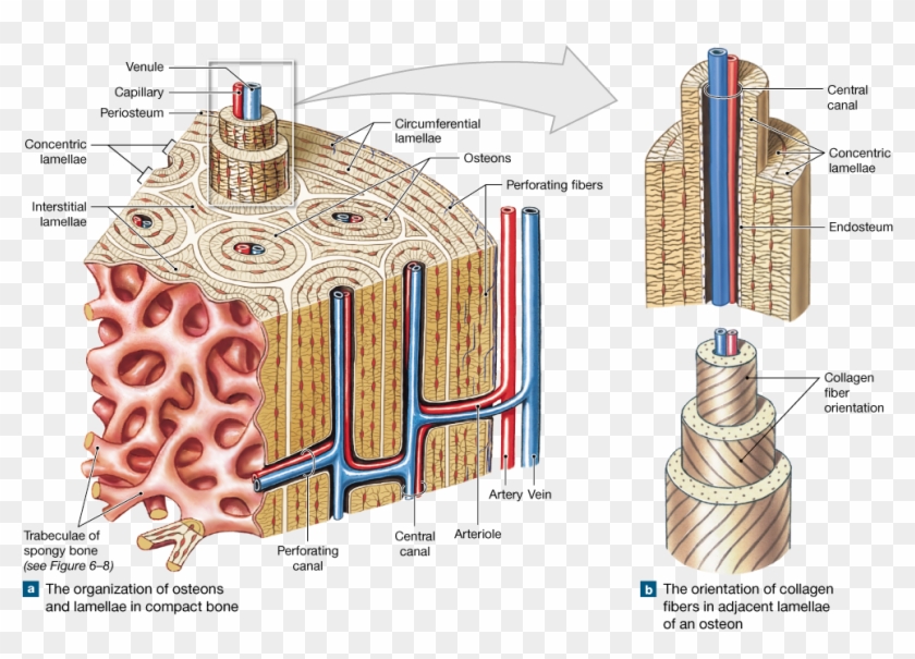
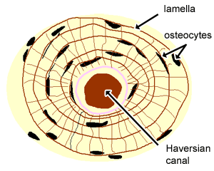
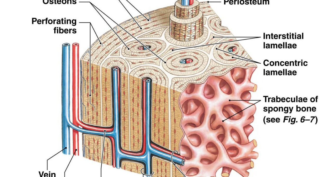
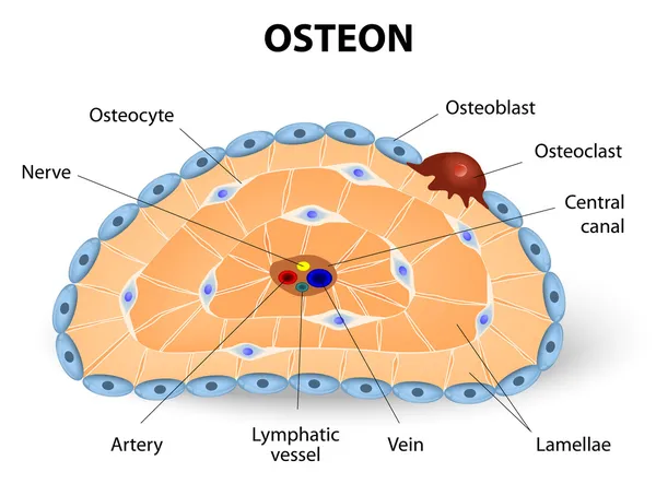
Here's a blown up view of an osteon. Another word for these osteons is the haversian system. So let's talk more about this haversian system. So each of these osteons looks like of like a cylinder and it has multiple concentric layers of bone, or sheets really, that wrap around each other to form this osteon. Each of these layers is called a ...
Osteons are roughly cylindrical structures that are typically between 0.25 mm and 0.35 mm in diameter. Their length is often hard to define, but estimates vary from several millimeters to around 1 centimeter. They are present in many bones of most mammals and some bird, reptile, and amphibian species. Contents 1 Structure 1.1 Drifting osteons
"Osteons" refers to systems of conduits (and associated structures) for small blood vessels and nerves that grow and run lengthwise in much compact bone of many vertebrates. Osteons respond to the...
In the diagram, where is the osteon?
A. Define osteon. B. Compare compact bone and spongy bone. C. A sample of bone has lamellae that are not arranged in osteons. Is the sample more likely from the epiphysis or from the diaphysis? Learning Outcome: Compare the structures and functions of compact bone and spongy bone.
80% (5 ratings) Central canal is also called harversian canal.It is a circular canal …. View the full answer. Transcribed image text: entify the structures of an osteon Part A Drag the labels onto the diagram to identify the structures of an osteon. Reset Help central canal JOIN lacuna lamella canac Submit Request Answer.
Mar 01, 2019 · Download scientific diagram | 3: Diagram of an osteon, the primary structural unit of bone, with the concentric locations of osteocytes shown (Vaughan et al.The microscopic structural unit of compact bone is called an osteon, or Haversian system. Each osteon is composed of concentric rings of calcified matrix called lamellae (singular = lamella ...
In the diagram, where is the osteon?.
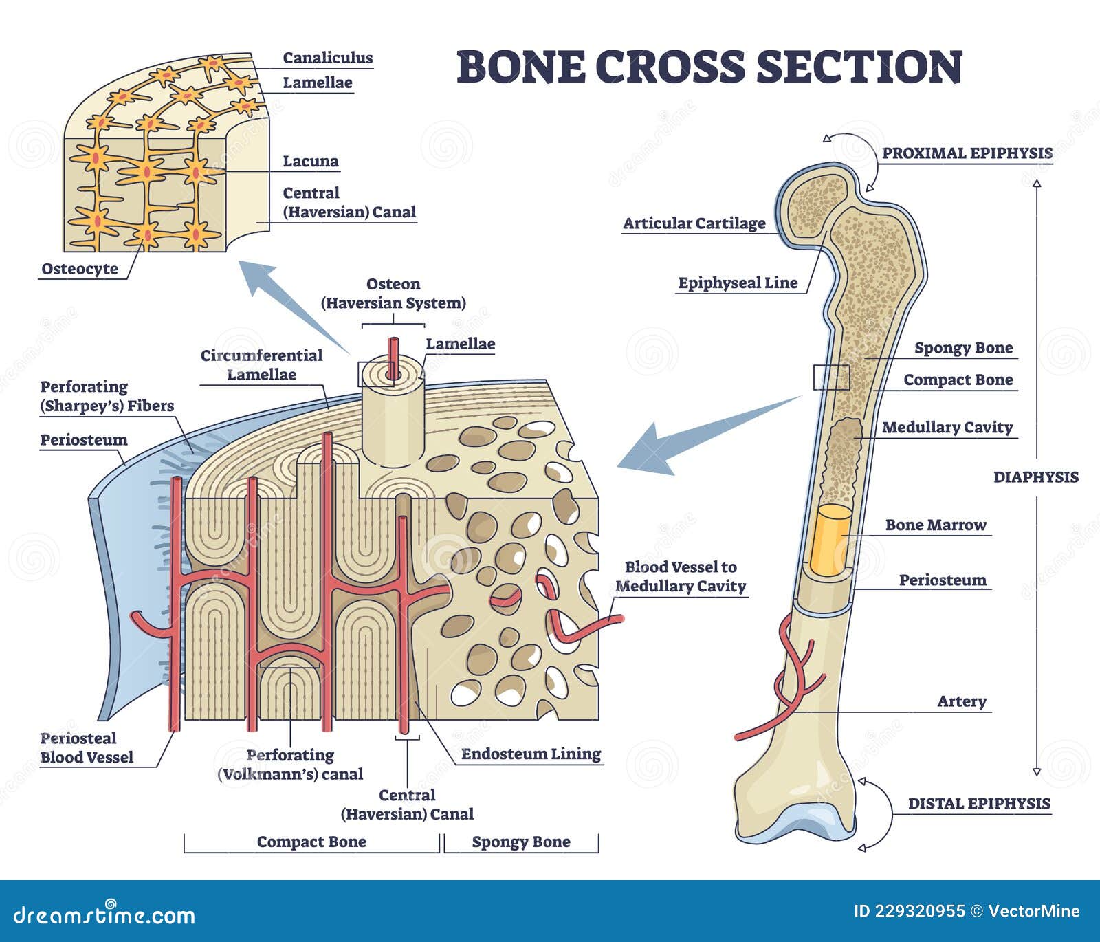
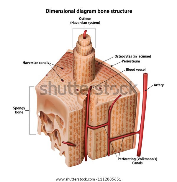
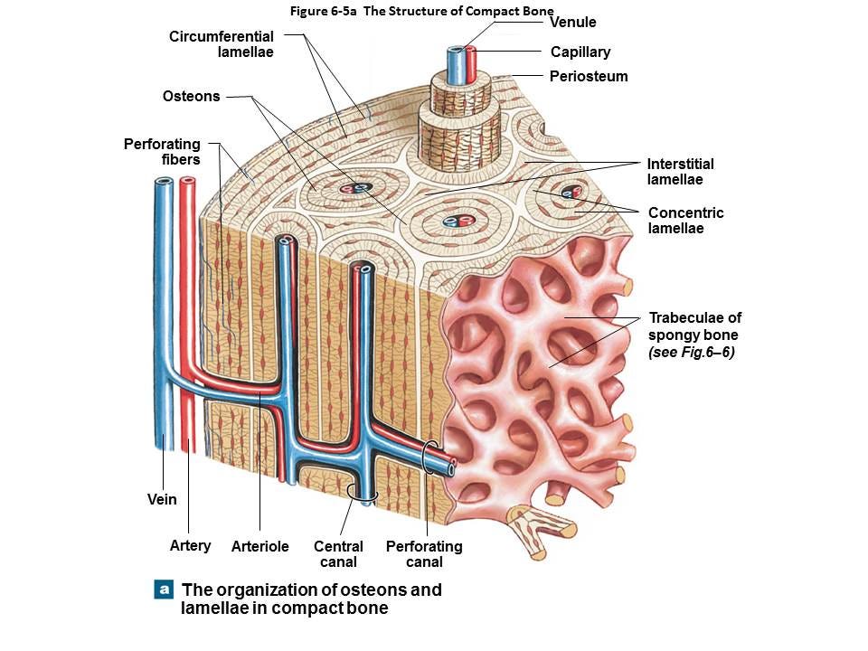
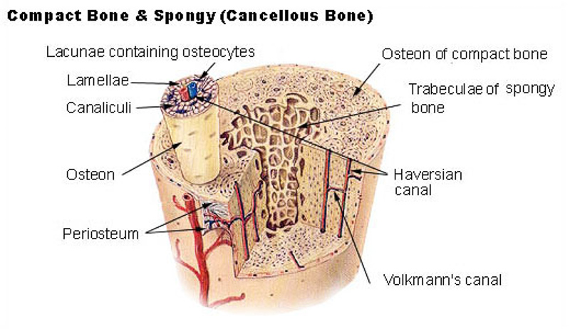
Compact Bone. Compact bone consists of closely packed osteons or haversian systems. The osteon consists of a central canal called the osteonic (haversian) canal, which is surrounded by concentric rings (lamellae) of matrix. Between the rings of matrix, the bone cells (osteocytes) are located in spaces called lacunae.
The basic first level structure of cortical bone are osteons. Trabecular bone is much more porous with porosity ranging anywhere from 50% to 90%. It is found in the end of long bones (see picture above), in vertebrae and in flat bones like the pelvis. Its basic first level structure is the trabeculae. III.
Indicated are Haversian canal with blood and lymphatic vessels, a nerve, and loose. Bodytomy provides a labeled diagram of the Haversian system to help you The terms 'Haversian system' or 'osteon' refer to the basic. osteon diagram TS. The diagram above shows a transverse view of an osteon ( Haversian system) - the basic unit of compact bone.
In the diagram, where is the osteon? B. In the diagram, where is the trabeculae? B. In the diagram, where is the zone of hypertrophic cartilage? A. In the diagram, this zone contains mostly dead chondrocytes surrounded by a calcified matrix. D. In the diagram, where is the zone of resting cartilage? A.
Osteons are several millimetres long and about 0.2 millimetre (0.008 inch) in diameter; they tend to run parallel to the long axis of a bone. Osteons are formations characteristic of mature bone and take shape during the process of bone remodeling, or renewal.
The terms 'Haversian system' or 'osteon' refer to the basic cylindrical-shaped structural unit of a compact bone, which in turn forms a substantial part of the structure of the long bones of the human body. The osteons are closely packed, with osteocytes lined up in concentric rings. This imparts a hard and dense texture to the compact bones.
Start studying Osteon Label. Learn vocabulary, terms, and more with flashcards, games, and other study tools.
100% (10 ratings) Answer There are two types of bone tissue : - Compact bone - Spongy bone Osteo …. View the full answer. Transcribed image text: Label the structural components of bone tissue in the diagram: 8 pts Osteon Compact bone Haveriana Canaliculus Lacuna Osteon Lamellae Spongy bone Spongy bone Osteocyte Compact bone.
central part of the osteon. provides passageway for bones, nerve, and blood supply. surrounded by rings of Lamellae. Canaliculi. Microscopic canals within lamellae that link osteocyte. Sets found in the same folder. head/cranium bones. 17 terms. l_dewey1.
1. Concentric rings made up of groups of hollow tubes of bone…. 1. center of osteon that contains nerves and blood vessels tha…. spaces within the osteons that contain osteocytes. 1. little channels that connect lacunae... 2. gap junctions allow…. Lamellae (3) 1.
Haversian system or osteon. This (Haversian system or osteon) is the structural unit of a compact bone matrix. They are the long cylindrical and branching structural unit that lies parallel to the long axis of the bone shaft. Each of the osteon or Haversian systems contains a centre canal or Haversian canal at the system's centre.
Figure 6.3.6 – Diagram of Compact Bone: (a) This cross-sectional view of compact bone shows several osteons, the basic structural unit of compact bone. (b) In this micrograph of the osteon, you can see the concentric lamellae around the central canals.
Compact bone contains parallel osteons, and spongy - osteons of ...
The osteon or haversian system /həˈvɜːr.ʒən/ (named for Clopton Havers) is the fundamental functional unit of much compact bone. Osteons are roughly cylindrical structures that are typically between 0.25 mm and 0.35 mm in diameter.[1] Their length is often hard to define,[2] but estimates vary from several millimeters[3] to around 1 centimeter.[1]

Structure and function of the haversian system explained with ...
About Press Copyright Contact us Creators Advertise Developers Terms Privacy Policy & Safety How YouTube works Test new features Press Copyright Contact us Creators ...

Tulang panjang compact bot osteon kerangka manusia, kombinasi ...
-were the marrow is located Basic unit of structure in compact bone comprised of Lamellae, central canal and osteocytes. Osteocyte OSTEON BONE DIAGRAM: AN OSTEON BY ASHLEY HOLMES Central Canal -part of the a bone cell, forms when an osteoblast becomes embedded in the matrix it has secreted Lamellae layers on bone tissue found in compact bone

Structure of bones | biology for majors ii
The diagram above shows a longitudinal view of an osteon. Some, mostly older, compact bone is remodelled to form these Haversian systems (or osteons ). The osteocytes sit in their lacunae in concentric rings around a central Haversian canal (which runs longitudinally).

Bone cells forming osteon. stock vector illustration of osteocyte ...
osteon the osteon or haversian system h ə ˈ v ɜːr ʒ ən named for clopton havers is the fundamental functional unit of much pact bone osteons are roughly cylindrical structures that are typically several millimeters long and around 0 2 mm in diameter they are present in many bones of most mammals and some bird reptile and amphibian species

3: diagram of an osteon, the primary structural unit of bone, with ...
This is an online quiz called Diagram of an Osteon. There is a printable worksheet available for download here so you can take the quiz with pen and paper. Your Skills & Rank. Total Points. 0. Get started! Today's Rank--0. Today 's Points. One of us! Game Points. 5. You need to get 100% to score the 5 points available.
Cartilage, bone & ossification: the histology guide
It can be found under the periosteum and in the diaphyses of long bones, where it provides support and protection. Diagram of Compact Bone (a) This cross-sectional view of compact bone shows the basic structural unit, the osteon. (b) In this micrograph of the osteon, you can clearly see the concentric lamellae and central canals. LM × 40.

Kanal haversian osteon bone compact bot kerangka manusia, tulang ...
UNIT 5/Chapter 5 - Review Guide - KEY. chapter_5_skeletal_system_review_guide_key.docx. File Size: 3324 kb. File Type: docx. Download File.

Bone osteon diagram | quizlet
Bodytomy provides a labeled diagram of the Haversian system to help you The terms 'Haversian system' or 'osteon' refer to the basic. osteon diagram TS. The diagram above shows a transverse view of an osteon ( Haversian system) - the basic unit of compact bone. diagram of haversian canal. This video was produced to help students of human anatomy.

Schematic diagram of compact and spongy bones. schematic diagram ...
The terms 'Haversian system' or 'osteon' refer to the basic cylindrical-shaped structural unit of a compact bone, which in turn forms a substantial part of the structure of the long bones of the human body. The osteons are closely packed, with osteocytes lined up in concentric rings. This imparts a hard and dense texture to the compact ...
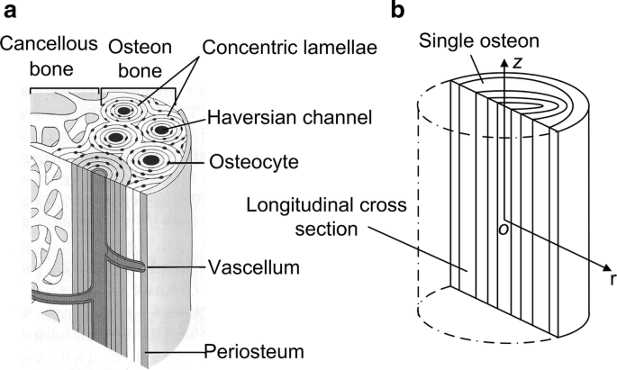
![a) Schematic representation of osteons in compact bone [2]. (b ...](https://www.researchgate.net/publication/343478647/figure/fig1/AS:921520050294786@1596718909634/a-Schematic-representation-of-osteons-in-compact-bone-2-b-Design-of-osteon.png)
A) schematic representation of osteons in compact bone [2]. (b ...
Definition. part of the osteon that is in the middle of the lamellae and contains blood vessels and nerves. These run longitudinally through the bone. Location. osteocytes. all bone cells belong to this group whether they build bone or destroy it. lacunae. in bone tissue, these are the little wells that the osteocytes sit in, chondrocytes sit ...

Test 2) osteons (haversian systems) diagram | quizlet

5 structure of the osteons of cortical bone (junqueira and ...

Ringkasan tulang | britannica

Anatomy of compact bone. compact bone tissue osteon diagram stock ...

Blood and interstitial flow in the hierarchical pore space ...

Sistem tubuh kita: rangka
Anatomy diagrams - labeled diagram images of things and processes

Bones structure and function -earn and extra 200-400 a week ...
Bone cross section and isolated anatomical detailed structure ...
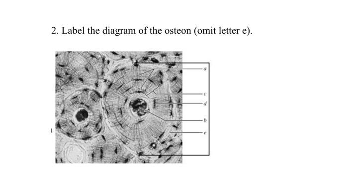
Solved 2. label the diagram of the osteon (omit letter e). | chegg.com
Vektor stok threedimensional diagram bone structure (tanpa royalti ...

Lab #1 - close-up of an osteon diagram | quizlet
Osteon stok vektor, ilustrasi osteon bebas royalti | depositphotos®
The systems of our body: skeletal | by sahasra pokkunuri ...

What is the structure and function of the compact bone? | socratic

Schematic diagram of compact and spongy bones. schematic diagram ...

Osteon model | anatomy models labeled, human body systems, human ...

Osteon - wikipedia

Osteon - wikipedia

Osteon 2 diagram | quizlet
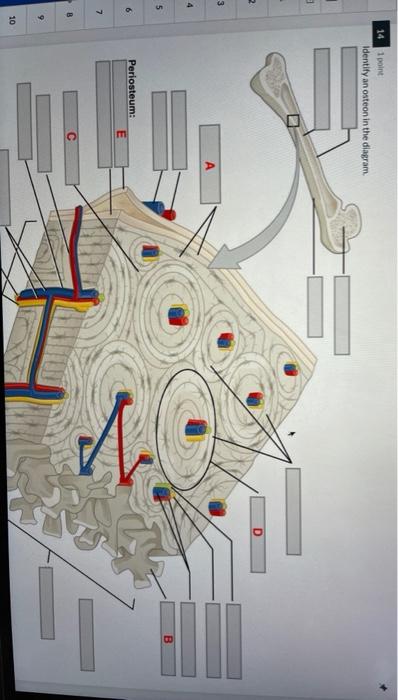
Solved 14 1 point identify an osteon in the diagram 2 3 a 4 ...

Ch.7 osteon diagram review diagram | quizlet

Tiara. on twitter: "cr ; zenius, gambar dari google" / twitter
Osteon - definition and synonyms of osteon in the german dictionary

File:osteon cross section.png - wikimedia commons
A multi-layered poroelastic slab model under cyclic loading for a ...

Bones: structure and types

Osteon and other label stuff flashcards | quizlet

6.3 bone structure – anatomy & physiology

Bone canaliculus - wikipedia
0 Response to "42 in the diagram, where is the osteon?"
Post a Comment