42 drag the labels onto the diagram to identify the types of epithelia.
layers of the epidermis..PNG - | Course Hero layers of the epidermis..PNG -. School Terra Community College. Course Title BIO 1230. Uploaded By tierrasarver50. Pages 1. Ratings 50% (4) 2 out of 4 people found this document helpful. This preview shows page 1 out of 1 page. Drag the labels onto the diagram to identify the tissues ... Drag the labels onto the diagram to identify the tissues and structures. Reset Help central cand matrix Group 2 lacuna Group 2 Group 2 osteocyte in lacuna Group 2 C chondrocyto Group 2 bono (osseous ... Cartilage and bone are two specialized types of connective tissues. Cartilage cells are .
Drag the labels onto the diagram to identify the layers of ... Drag the labels onto the diagram to identify the various types of cutaneous receptors. Reset Help G Free nerve endings (pain temperature) Lamellar corpuscle (deep pressure) Dermis Tactile corpuscle (touch, light pressure) Epidermis Hair follicle...
Drag the labels onto the diagram to identify the types of epithelia.
Ch 13 lab Map, Ch 12 lab map, CH 11 Lab MAP, Ch 10 ... - Quizlet Drag the labels onto the diagram to identify the major skeletal muscles, anterior view. look at pic Cells located in each seminiferous tubule which function to provide a microenvironment that supports spermatogenesis are called nurse cells Drag the labels onto the diagram to identify the parts of the male reproductive system. look at pic Simple Columnar Epithelium: A Labeled Diagram and ... Epithelium is a tissue that lines the internal surface of the body, as well as the internal organs. Simple epithelium is one of the types of epithelium that is divided into simple columnar epithelium, simple squamous epithelium, and simple cuboidal epithelium. Bodytomy provides a labeled diagram to help you understand the structure and function of simple columnar epithelium. Part A Drag the labels onto the diagram to identify the ... Part A Drag the labels onto the diagram to identify the structures of the neuron. ANSWER: cutaneous membrane. serous membrane. mucous membrane. synovial membrane. peritoneal membrane. fibrocartilaginous membranes serous membranes cutaneous membranes mucous membranes synovial membranes
Drag the labels onto the diagram to identify the types of epithelia.. Solved Drag the labels onto the diagram to identity the ... Drag the labels onto the diagram to identity the types of epithelia. Question : Drag the labels onto the diagram to identity the types of epithelia. This problem has been solved! Drag The Labels Onto The Diagram To Identify The ... 32 Label The Structures Of The Plasma Membrane And Cytoskeleton ... Drag The Labels Onto The Diagram To Identify The Structures Or Steps In ... Drag The Labels Onto The Diagram To Identify Structural Features ... Animal Tissues - Epithelium, Connective Tissues | PMF IAS UPSC. Mastering A&P Exam 1 Flashcards & Practice Test - Quizlet The framework or stroma of organs such as the spleen, liver, and lymph nodes is made up of ________ tissue. collagen, reticular, and elastic. The three types of protein fibers in connective tissue are. (week three assignment one) #6. Drag the labels onto the diagram to identify the types of connective tissue proper. a&p lab 6 hw Flashcards - Quizlet a&p lab 6 hw. Drag the labels onto the diagram to identify the classes of epithelia based on number of cell layers and cell shape. (figure 6.2) This tissue type is a covering and lining tissue. It also includes glands. Nice work!
4.2 Epithelial Tissue - Anatomy & Physiology Both simple and pseudostratified columnar epithelia are heterogeneous epithelia because they include additional types of cells interspersed among the epithelial cells. For example, a goblet cell is a mucous-secreting unicellular gland interspersed between the columnar epithelial cells of a mucous membrane ( Figure 4.2.3 ). Drag the labels onto the diagram to identify the blood ... Drag the labels onto the diagram to identify the types of epithelia. Posted 13 days ago. Q: Drag the labels onto the diagram to identify the stages in which the lagging strand is synthesized. Drag the labels onto the diagram to identify the stages in which the lagging strand is synthesized. Solved Art-labeling Activity: Classifying Epithelia Drag the ... Art-labeling Activity: Classifying Epithelia Drag the labels onto the diagram to identify the types of epithelia. Reset Help Stratified Simple squamous epithelium Strated columna epithelum Simple columnas epithelium Cuboidal Stratified squamous epithelum Squamous Strated cuboidal opithelium Simple cuboidal epithelium Simple Columnar Adel Noa - Lire En Ligne Drag The Labels Onto The Diagram To Identify The Types Of Epithelia Drag each label into the appropriate position to identify how each theoretical condition would alter body function. To answer the question you need Reflexes or reflex actions are involuntary almost instantaneous movements in response to a specific stimulus.
Epithelial Tissue - Anatomy and Physiology The different types of epithelial tissues are characterized by their cellular shapes and arrangements: squamous, cuboidal, or columnar epithelia. Single cell layers form simple epithelia, whereas stacked cells form stratified epithelia. Very few capillaries penetrate these tissues. Drag the labels onto the diagram to identify the types of ... Q:Drag the labels onto the diagram to identify the types of epithelia Q:Contrary to popular belief, psychologists now believe that catharsis A-is a very healthy and necessary method of expres Q:19. you are interviewing an employee of a company to find out about the job, the nature of the work, employment setting Drag the labels onto the diagram to identify the types of ... Drag the labels onto the diagram to identify the classes of epithelia based on number of cell layers and cell shape. Posted 12 days ago Q: Part A Drag the labels onto the diagram to identify the bones and markings of the skull. Solved Art-labeling Activity: Classifying epithelia Drag ... Art-labeling Activity: Classifying epithelia Drag the labels onto the diagram to identify the types of epithelia. Reset pihem Stratified Simplo columna opithelium Simple Squamous Stratified columnar epithelium Stratified squamous epithelium Simple squamous epithelium Columnar
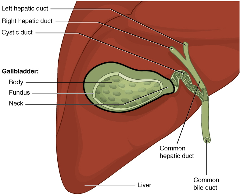
Gallbladder and Biliary Tract: Anatomy | Concise Medical ...
Drag the labels onto the diagram to identify the ... Drag the labels onto the diagram to identify the types of epithelia. Posted 7 days ago. Q: Drag the labels onto the diagram to identify the stages in which the lagging strand is synthesized. Drag the labels onto the diagram to identify the stages in which the lagging strand is synthesized.
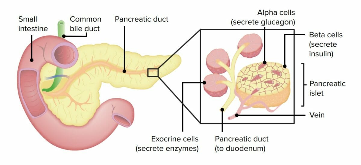
Gastrointestinal Secretions | Concise Medical Knowledge
API Lab Homework 2 - 10/7/2016 APILabHomework2 ... 10/7/2016 API Lab Homework 2 1/9 API Lab Homework 2 Due: 11:59pm on Friday, October 14, 2016 You will receive no credit for items you complete after the assignment is due. Grading Policy Artlabeling Activity: Anatomy of a Model Cell, Part 1 Learning Goal: To learn the components of a model cell. Label the components of a model cell. Part A Drag the labels onto the diagram to identify the ...
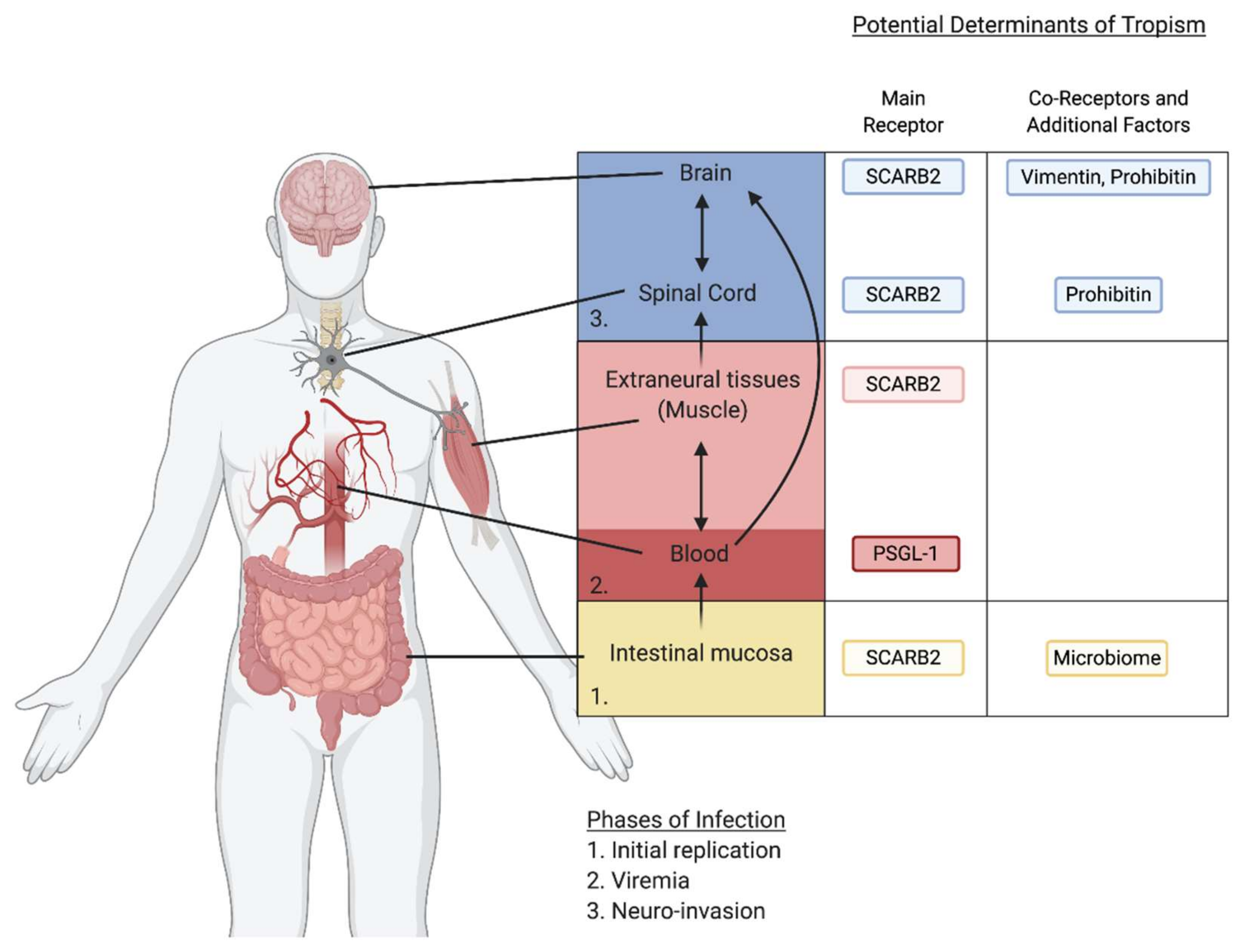
Viruses | Free Full-Text | Return of the Neurotropic ...
HW 2.pdf - HW 2 Due: 11:59pm on Friday ... - Course Hero Label the types of cell junctions. Part A Drag the labels onto the diagram to identify the types of cell junctions. ANSWER: Correct Art-labeling Activity: The Polarity of Epithelial Cells Identify the structures in epithelial cells. Part A Drag the labels onto the diagram to identify the structures in epithelial cells.

Dielectric characterization of bioparticles via ...
Drag The Labels Onto The Diagram To Identify The Stages Of ... Drag the labels onto the diagram to identify the stages of the life cycle. Drag the labels onto the diagram to identify the stages of the cell cycle.. Drag the with resolution 2232px x 1472px. Drag the pink labels.
Anatomy and Physiology Lab I” on OpenALG
Solved Drag the labels onto the diagram to identify the ... Experts are tested by Chegg as specialists in their subject area. We review their content and use your feedback to keep the quality high. Transcribed image text: Drag the labels onto the diagram to identify the types of epithelia.

Leishmania Hijacks microRNA Import-Export Machinery of ...
LAB A&P Flashcards | Quizlet Drag the labels onto the diagram to identify the types of epithelia. look at the picture Exfoliative cytology involves the removal of epithelial cells for examination.
Dissertation
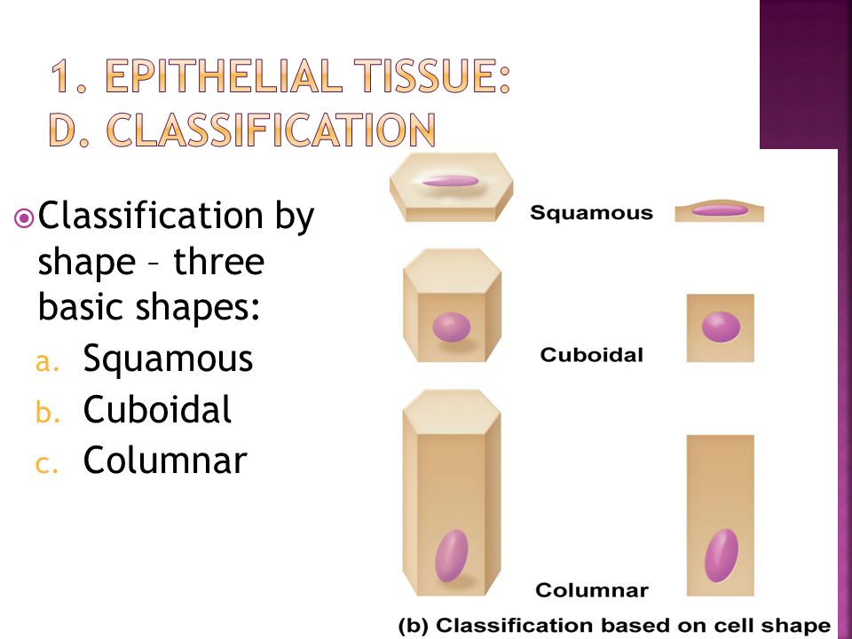
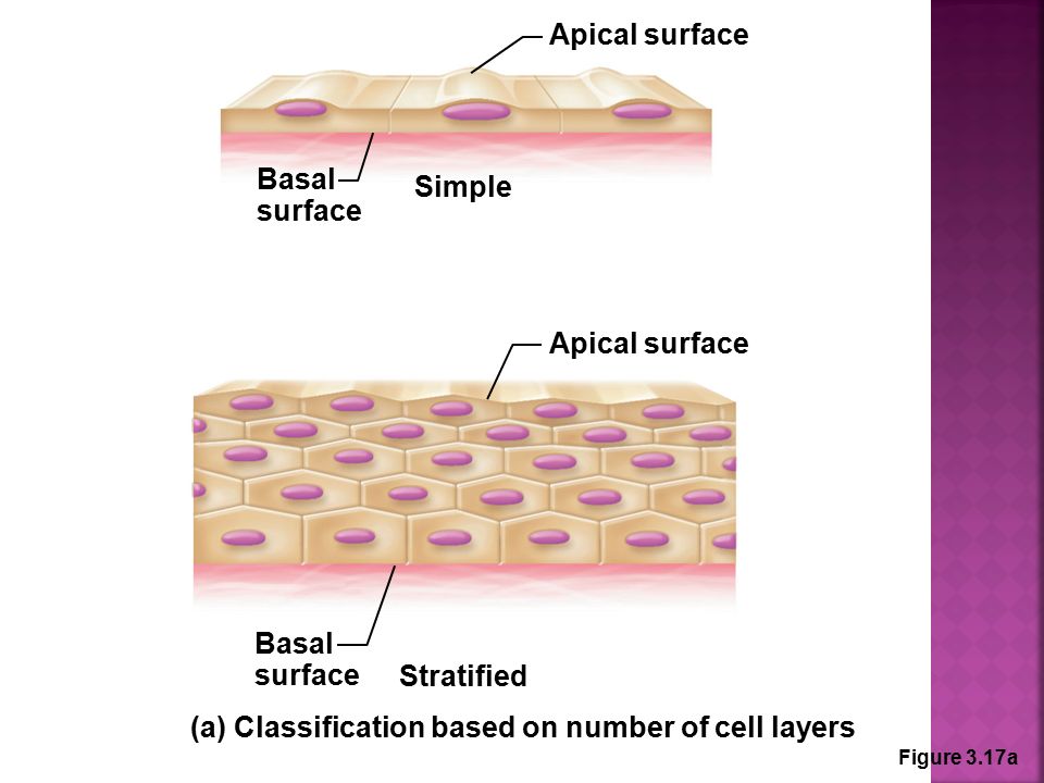
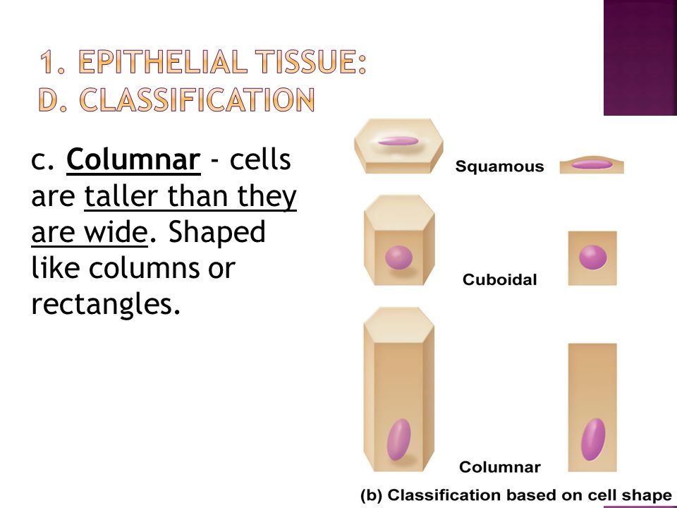
PDF Marieb HA8 chapter 4 - Pearson Classification of Epithelia Many kinds of epithelia exist in the body. Two features are used to classify and name epithelia: the number of cell layers and the shape of the cells (Figure 4.2). The terms simple and stratified describe the number of cell layers in an epithelium (Figure 4.2a). t Simple epithelia contain a single layer of cells ...

Drag The Labels To Identify The Forms Of Immunity. - FORM.EAD ...
Solved Part A Drag the labels onto the diagram to identify ... Transcribed image text: Part A Drag the labels onto the diagram to identify the classes of epithelia based on number of cell layers and cell shape.
![Expert Answer] Drag labels to the appropriate locations in ...](https://us-static.z-dn.net/files/dd9/363dc6f0791b99410cbee879fdbd9356.png)
Expert Answer] Drag labels to the appropriate locations in ...
Ch 5-7 Lab A&P Mastering Flashcards - Quizlet Drag the labels onto the diagram to identify the types of epithelia. look at the picture Exfoliative cytology involves the removal of epithelial cells for examination. Which of the following is NOT a clinical application of exfoliative cytology? examination of cardiac muscle cells after a heart attack
Answered: Drag the labels to the appropriate… | bartleby
Solved: Drag The Labels Onto The Diagram To Identify The S A20 Visually enhanced, image enriched topic search for Solved: Drag The Labels Onto The Diagram To Identify The S A20.
Characterization of TRP Ion Channels in Cardiac Muscle A ...
Solved Art-labeling Activity: Classifying Epithelia ... Experts are tested by Chegg as specialists in their subject area. We review their content and use your feedback to keep the quality high. Transcribed image text: Art-labeling Activity: Classifying Epithelia Identify the types of epithelia. Drag the lables onto the diagram to identify the types of epithelia.
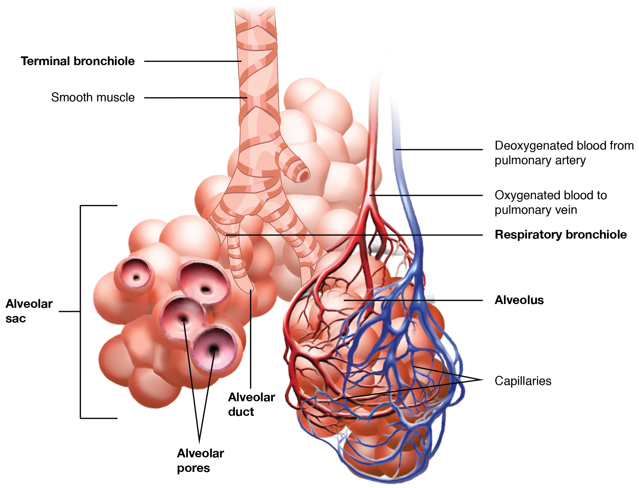
Respiratory System - Medicine LibreTexts
Part A Drag the labels onto the diagram to identify the ... Part A Drag the labels onto the diagram to identify the structures of the neuron. ANSWER: cutaneous membrane. serous membrane. mucous membrane. synovial membrane. peritoneal membrane. fibrocartilaginous membranes serous membranes cutaneous membranes mucous membranes synovial membranes
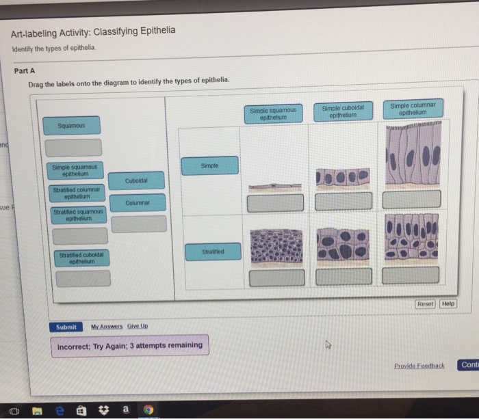
Solved Art-labeling Activity: Classifying Epithelia | Chegg.com
Simple Columnar Epithelium: A Labeled Diagram and ... Epithelium is a tissue that lines the internal surface of the body, as well as the internal organs. Simple epithelium is one of the types of epithelium that is divided into simple columnar epithelium, simple squamous epithelium, and simple cuboidal epithelium. Bodytomy provides a labeled diagram to help you understand the structure and function of simple columnar epithelium.

Oat Fiber Modulates Hepatic Circadian Clock via Promoting Gut ...
Ch 13 lab Map, Ch 12 lab map, CH 11 Lab MAP, Ch 10 ... - Quizlet Drag the labels onto the diagram to identify the major skeletal muscles, anterior view. look at pic Cells located in each seminiferous tubule which function to provide a microenvironment that supports spermatogenesis are called nurse cells Drag the labels onto the diagram to identify the parts of the male reproductive system. look at pic
Solved] Drag the labels onto the diagram to identify the ...
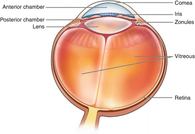
The Eye and Its Anatomical Considerations | SpringerLink

Ch 18_lecture_presentation
Answered: ne labels onto he diagram Турes… | bartleby
Define tissue Describe the germ tissue layers, their ...

Peterson, Michael / Book Images
Define tissue Describe the germ tissue layers, their ...
Tissues
Mam Phys Exam 2 Flashcards - Cram.com

Hyaline cartilage structure and biochemical composition ...

Ch 5-7 Lab A&P Mastering Flashcards | Quizlet

Skin Disorders | Human Anatomy Quiz - Quizizz

Ch 5-7 Lab A&P Mastering Flashcards | Quizlet

4.2 Epithelial Tissue – Anatomy & Physiology

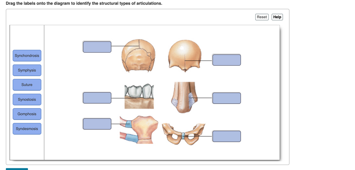
11.4 Identify the skeletal muscles and give their origins ...
WALL OF THE HEART AND CARDIAC VALVES
Define tissue Describe the germ tissue layers, their ...

Trajectories of the best and worst performers on the immune ...
This electronic thesis or dissertation has been downloaded ...

Stalling interkinetic nuclear migration in curved ...
Neural Control of Human Movement
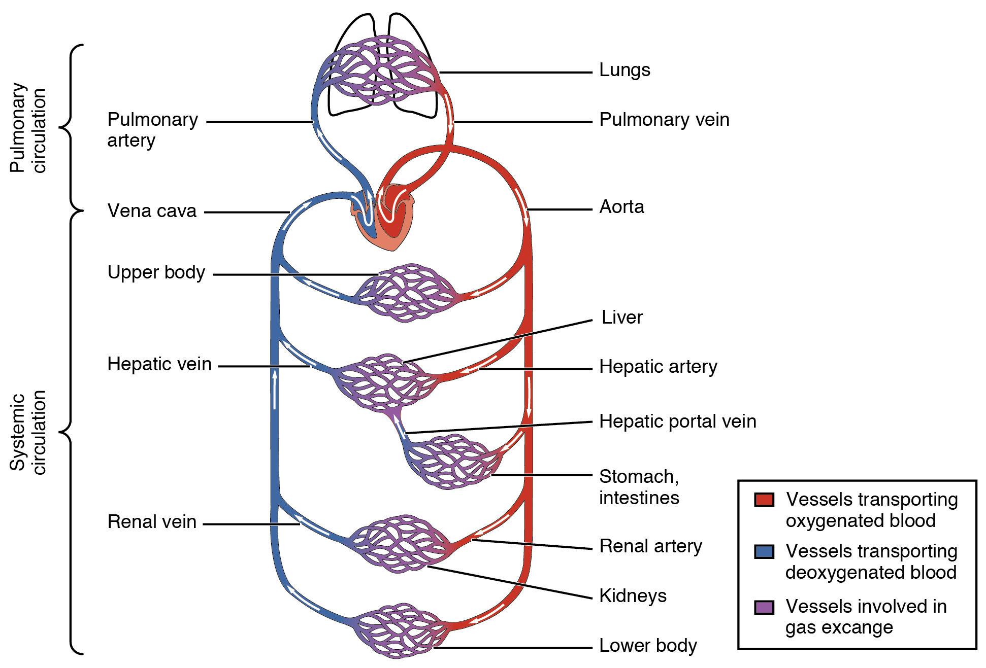
Structure and Function of Blood Vessels – Anatomy and Physiology

Epithelial Tissue | Anatomy and Physiology I
Pathway Logic Guided Tour: STM8

Glycosylation in HIV-1 envelope glycoprotein and its ...

Taste receptor - Wikipedia

ANAT 3010 Lecture Notes - Winter 2020, Lecture 5 - Mandibular ...
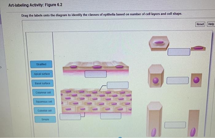
Solved Art-labeling Activity: Figure 6.2 Drag the labels ...
0 Response to "42 drag the labels onto the diagram to identify the types of epithelia."
Post a Comment