45 pupillary light reflex diagram
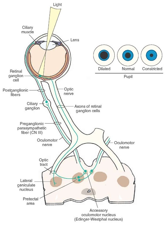
Pupillary Light Reflex Pathway. Pupillary Light Reflex Pathway, is a reflex that controls the diameter of the pupil, in response to the intensity (luminance) of light that falls on the retina of the eye, thereby assisting in adaptation to various levels of darkness and light, in addition to retinal sensitivity. pupillary light reflex pupil size change in response to amt of light to maintain luminance of retina; in addation to those linked w/A; 4 neuron pathway direct response
http://www.handwrittenturorials.com - This tutorial is the second in a series of tutorials on the reflexes of the brainstem. This video covers the function a...

Pupillary light reflex diagram
Schematic Diagram Of The Pupil Light Reflex Showing Negative Pupil Frontiers Functional Organization Of The Sympathetic Pathways 15 2 Autonomic Reflexes And Homeostasis Anatomy And Physiology Solved The Light Reflex Figure 5 Is A Pupillary Reflex Reflexes Clasifications And Functions Ocular Movements Visual Reflexes Medatrio ... Pupillary light reflex. The pupillary light reflex is an autonomic reflex that constricts the pupil in response to light, thereby adjusting the amount of light that reaches the retina 1).Pupillary constriction occurs via innervation of the iris sphincter muscle, which is controlled by the parasympathetic system 2).. Testing of the pupillary light reflex is useful to identify a relative ... …the best-known reflex is the pupillary light reflex. If a light is flashed near one eye, the pupils of both eyes contract. Light is the stimulus; ...
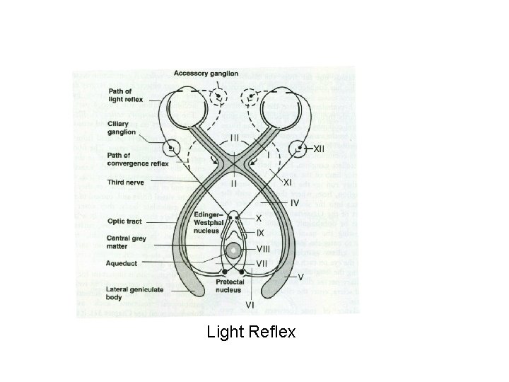
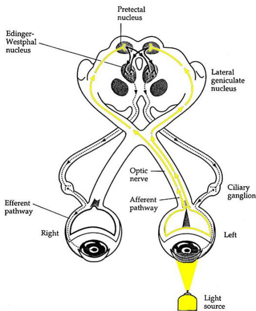
Pupillary light reflex diagram. (B on Fig. 1 - Pupillary Light Reflex Diagram) 1. Blind in left eye. 2. Direct pupillary reflex absent on left. 3. Consensual pupillary reflex absent on the right when light shined in left eye. 4. Near response present both eyes. C. Blindness due to occlusion of right posterior cerebral artery or middle cerebral artery. (C on Fig. 2 - Near ... The pupillary light reflex two main parts: an afferent limb and an efferent limb. The diagram below shows the neuroanatomical pathways of the pupillary light ... Download scientific diagram | Schematic drawing of the pupillary light reflex pathway. By way of the optic tract the afferent pathway (1) of the pupillary system projects to the dorsal midbrain. (B on Fig. 1 - Pupillary Light Reflex Diagram) 1. Blind in left eye. 2. Direct pupillary reflex absent on left. 3. Consensual pupillary reflex absent on the right when light shined in left eye. 4. Near response present both eyes. C. Blindness due to occlusion of right posterior cerebral artery or middle cerebral artery. (C on Fig. 2 - Near ...
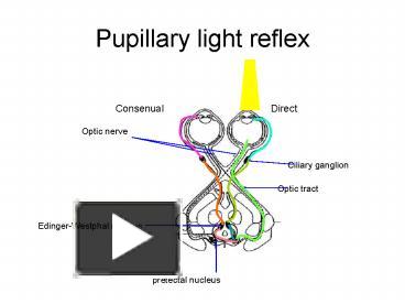
pupillary light reflex · The reflex is consensual: Normally light that is directed in one eye produces pupil constriction in both eyes. · The direct response is ... Diagram shows pathway of the pupillary light reflex. This is a schematic diagram of the pupillary light reflex. The afferent limb originates in the retinal photoreceptors, which convert light energy to a neural signal. Pupillary information is conveyed from the eye to the brain by the melanopsin-expressing retinal ganglion cells, and their ... Portable and easy to use, Pupillary Light Reflex study sets help you review the information and examples you need to succeed, in the time you have available. Use your time efficiently and maximize your retention of key facts and definitions with study sets created by other students studying Pupillary Light Reflex. The pupillary light reflex (PLR) or photopupillary reflex is a reflex that controls the diameter of the pupil, in response to the intensity of light that falls on the retinal ganglion cells of the retina in the back of the eye, thereby assisting in adaptation of vision to various levels of lightness/darkness. A greater intensity of light causes the pupil to constrict (miosis/myosis; thereby ...
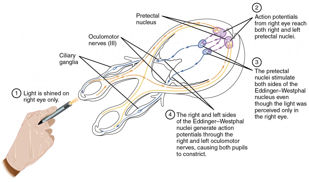
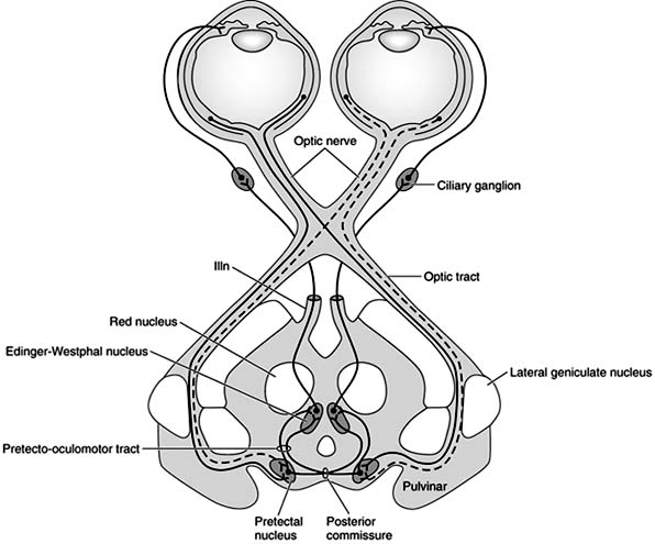
Figure 2 Schematic diagram of the pathway for the pupillary light reflex. taining homeostasis. Autonomic reflex control occurs at different levels. 1. Local reflexes such as bladder contraction, where pelvic sensory nerves form the afferent limb, relaying in the spinal cord at S2-S4 with In afferent pupillary defects as in optic nerve lesions, the pupil does not react to light reflex, but near reflex will be present. Interestingly, even though the same neural machinery is involved in both the accommodation reflex and the pupillary light reflex, certain conditions demonstrate a dissociation between the two pathways; this is ... Pupillary Light Reflex Clinical Exam Anatomy Pathway Sections Further Reading . Clinical Exam . ... Stimulation of the contralateral CN III parasympathetic nucleus results in this diagram from optic nerve transmission which does not cross over at the optic chiasm (25% in the dog) but does so at the pretectal nucleus. ... Mar 18, 2008 — Figure 3 Schematic drawing of the pupillary light reflex pathway (figure modified from Wilhelm21,52). Afferent fibers from the retina travel ...
The pupillary light reflex is an autonomic reflex that constricts the pupil in response to light, thereby adjusting the amount of light that reaches the retina.Pupillary constriction occurs via innervation of the iris sphincter muscle, which is controlled by the parasympathetic system .. Pathway: Afferent pupillary fibers start at the retinal ganglion cell layer and then travel through the ...
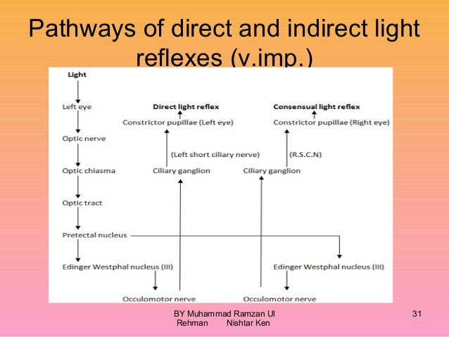
14.31 Pupillary Light Reflex. The pupillary light reflex requires CN II, CN III, and central brain stem connections. Light shined in one eye stimulates retinal photoreceptors, and subsequently retinal ganglion cells, whose axons travel through the optic nerve, chiasm, and tract to terminate in the pretectum (pretectal nucleus).

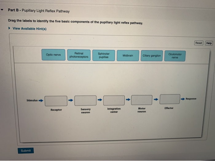
The pupillary light reflexes rely on a reflex pathway with the optic nerve as the sensory nerve, the oculomotor nerve as the motor nerve and the midbrain as the processing centre. How to Elicit Shine a light into each pupil.
The pupillary light reflex neural circuit: The pathway controlling pupillary light reflex (Figure 7.3) involves the. retina, optic nerve, optic chiasm, and the optic tract fibers that join the ; brachium of the superior colliculus, which terminate in the ; pretectal area of the midbrain, which sends most of its axons bilaterally in the posterior commissure to terminate in the
http://www.usmlesucess.net A USMLE tutorial covering the basics of the pupillary light reflex. Download your FREE copy of the Step 1 & Step 2 CK BIBLES at ...

The light reflex refers to the constriction that happens in both pupils when light shines through either eye. 2. Facts about The Human Eye As the window of the heart, eyes contains some facts that people had few noticed. Blind people with eyes also can feel the difference between light and darkness. There are individual cells in the eyes which ...
Concept Questions: Patellar Reflex and Pupillary Reflex Stations 1.In the following diagram, label as many as possible of the components involved in the patellar reflex: 2. In the following diagram, label the nervous system components involved in both the direct (same eye) and consensual (opposite eye) pupillary light reflex:
The pupillary light reflex is a reflex that controls the diameter of the pupil when it is exposed to varying intensities of light. This allows the eyes to adjust in response to bright or dim lights. Walk into any room and switch on the light; everything seems perfectly in its place.

The pupillary light reflex two main parts: an afferent limb and an efferent limb. The diagram below shows the neuroanatomical pathways of the pupillary light reflex. The details of the pathway are detailed below the diagram. Afferent Pathway of Pupillary Light Reflex (solid yellow above): Light enters the pupil and stimulates the retina ...
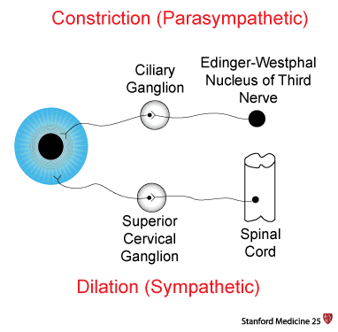
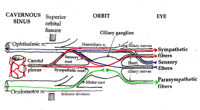
The physiology behind a "normal" pupillary constriction is a balance between the sympathetic and parasympathetic nervous systems. Parasympathetic innervation leads to pupillary constriction. A circular muscle called the sphincter pupillae accomplishes this task. The fibers of the sphincter pupillae encompass the pupil.
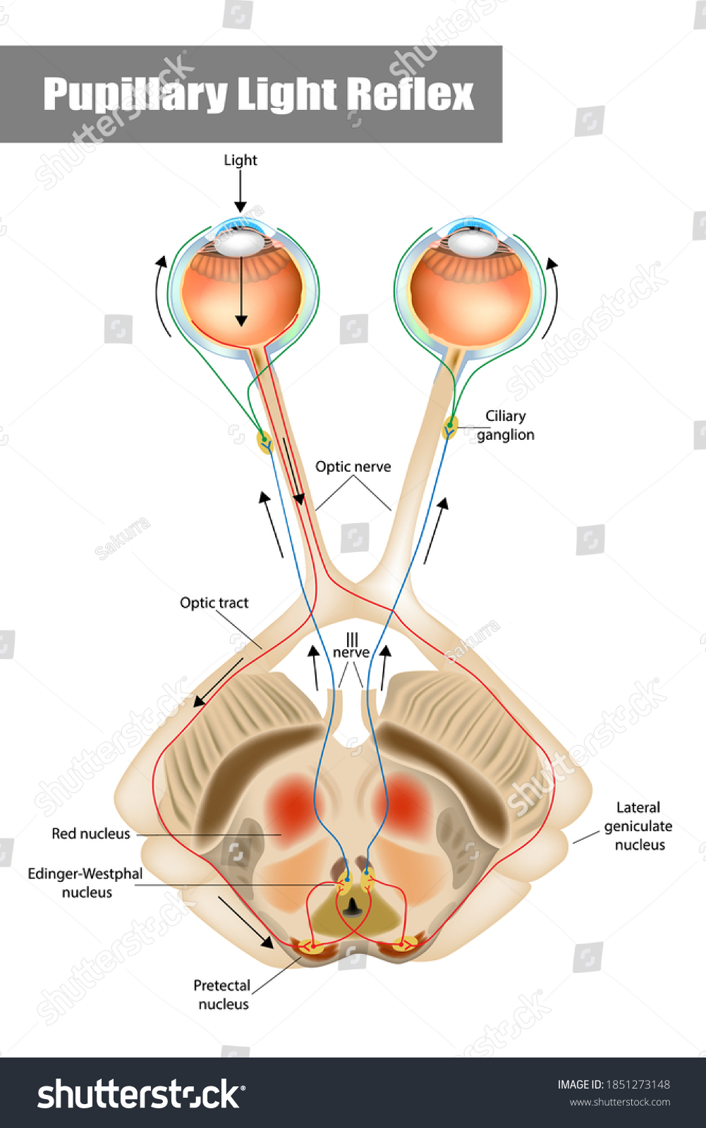
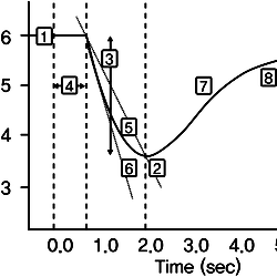
Figure P4.2 displays the block diagram of a simplified, linear closed-loop model used to characterize the pupillary light reflex. The input is AL (change in light intensity) and the output is ΔΑ (change in pupil area), and the gains Gi and G2 are nonnegative. (a) Derive expressions for the open- and closed-loop transfer functions of this ...
Download scientific diagram | Schematic diagram of the pupil light reflex showing negative feedback loop. from publication: Spontaneous oscillations in a nonlinear delayed-feedback shunting model ...

The pupil reflex. The amount of light entering the eye is controlled by a reflex action. The size of the pupil changes in response to bright or dim light. This is controlled by the muscles of the ...

ANATOMICAL PATHWAY OF PUPILLARY LIGHT REFLEX 1. Draw a labeled diagram of pupillary reflex pathway. (5) J2012 2. Pupillary pathways with diagram.(5) J2016 (1) Pupillary fibers in the nerve (2) Chiasmal decussation (3) Uncrossed fibers (4) Crossed fibers (5) Brachium of the superior colliculus with B/L decussation
The pupillary light reflex constricts the pupil in response to light, and pupillary constriction is achieved through the innervation of the iris sphincter muscle. Anatomy and Physiology Light travels through the cornea, anterior chamber, pupil, lens, and the posterior chamber, eventually reaching the retina.

…the best-known reflex is the pupillary light reflex. If a light is flashed near one eye, the pupils of both eyes contract. Light is the stimulus; ...

Pupillary light reflex. The pupillary light reflex is an autonomic reflex that constricts the pupil in response to light, thereby adjusting the amount of light that reaches the retina 1).Pupillary constriction occurs via innervation of the iris sphincter muscle, which is controlled by the parasympathetic system 2).. Testing of the pupillary light reflex is useful to identify a relative ...

Schematic Diagram Of The Pupil Light Reflex Showing Negative Pupil Frontiers Functional Organization Of The Sympathetic Pathways 15 2 Autonomic Reflexes And Homeostasis Anatomy And Physiology Solved The Light Reflex Figure 5 Is A Pupillary Reflex Reflexes Clasifications And Functions Ocular Movements Visual Reflexes Medatrio ...


0 Response to "45 pupillary light reflex diagram"
Post a Comment