40 neuron diagram and functions
Neurons, also known as nerve cells, are essentially the cells that make up the brain and the nervous system. Neurons do not touch each other, but where one neuron comes close to another neuron, a synapse is formed between the two. Behance Discovery - Alexey Kashpersky
A neuron is responsible for receiving sensory input, sending motor commands to your muscles, and transforming and relaying the electrical signals throughout these processes. Every neuron has one axon that connects it with other neurons or with muscle or gland cells.
Scientists May Have Unlocked Function of Mysterious Structure Found on Neurons in the Brain. Unusual clusters on neurons are calcium-signaling "hotspots" that activate gene transcription, allowing neurons to produce crucial proteins. For 30 years, mysterious clusters of proteins found on the cell body of neurons in the hippocampus, a part ...

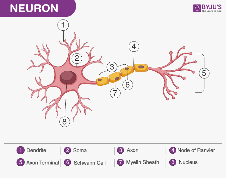
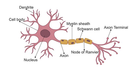
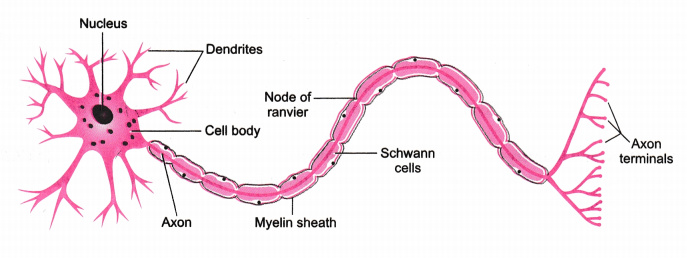
Neuron diagram and functions
There are more than 100 billion interneurons in the human body, which makes them the most abundant of the three major neuron types (along with sensory and motor neurons). ... Function, Structure ...
These neurons exist and function between sensory & motor neurons in the CNS. Impulses The nervous impulse is called 'action potential' as it is an electric signal produced by a neuron when it gets stimulated by external factors. A stimulus is a change in the environment with sufficient strength to initiate a response.
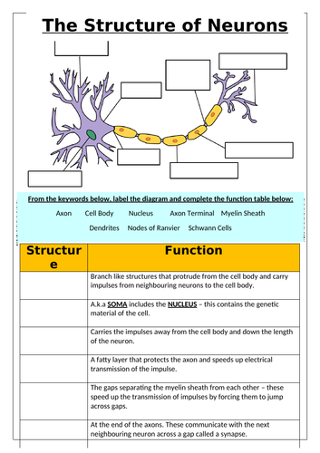
The functions of neurons are mentioned below: Neurons are the nerve cells that send and receive signals from the human brain. They have specific projections called axons to transmit electrical and chemical signals. In chemical synapses, electrical signals are converted into chemical signals or neurotransmitters.
Neuron diagram and functions.
The nervous system is a network of neurons whose main feature is to generate, modulate and transmit information between all the different parts of the human body. This property enables many important functions of the nervous system, such as regulation of vital body functions ( heartbeat, breathing, digestion), sensation and body movements.
The functions of synapses depend on where they are found. The three forms based on their location are: axodendritic - axon of one neuron and the dendrite of the next adjacent neuron. This is the most common form of synapse that dominates the nervous system; axosomatic - axon of one neuron and the cell body of the other neuron; axoaxonic - between two axons.
A neuron (nerve cell) is a specialized cell that conveys electrochemical impulses throughout the body. The cytology of a neuron facilitates the transmission of either: 'top-down' information from the brain to the periphery, via efferent neurons (e.g. to permit locomotion) (efferent neurons) or,
The functions of the PNS are commonly described as the "rest and digest" response, since it is involved in slowing down the heart rate, relaxing the sphincter muscles in the gastrointestinal and urinary tracts and increasing intestinal and gland activity.
Cell Membrane Neuron. The membrane with its mosaic of proteins is responsible for many important functions: keeping certain ions and small molecules out of the cell and letting others in, A resting (non-signaling) neuron has a voltage across its membrane called the resting membrane potential, or simply the resting potential.
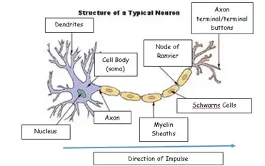
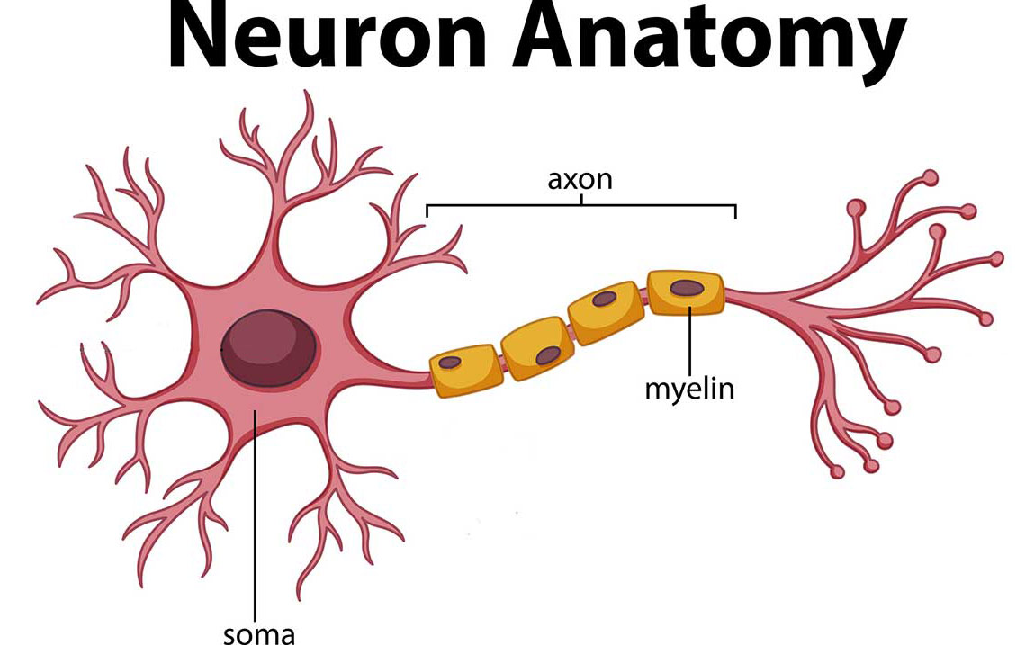
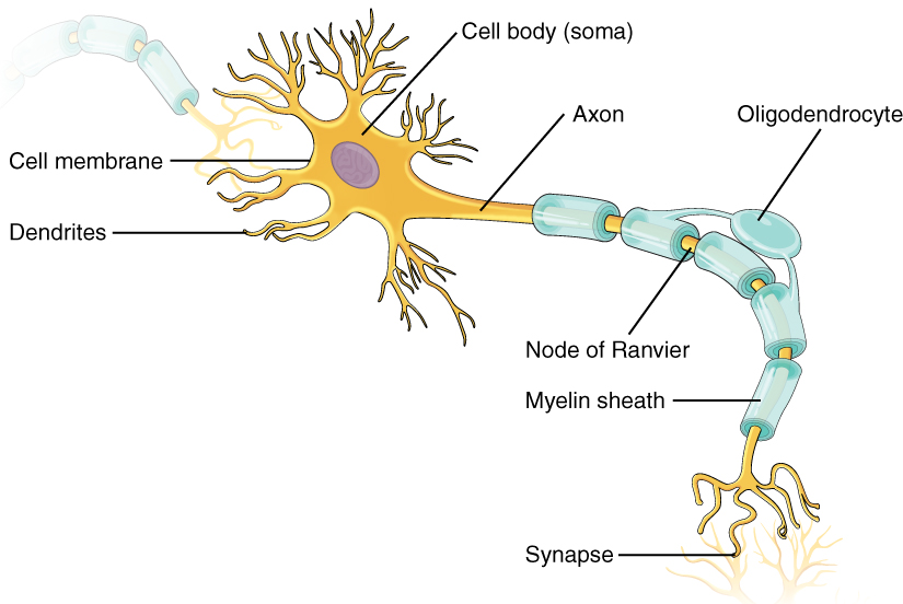
Below is a diagram of the parts of a neuron. Myelin insulates the neuron like plastic insulates electrical wires. Neurons send signals using electricity, so the myelin helps to keep the signal...
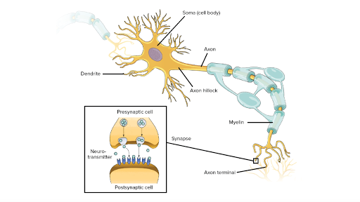
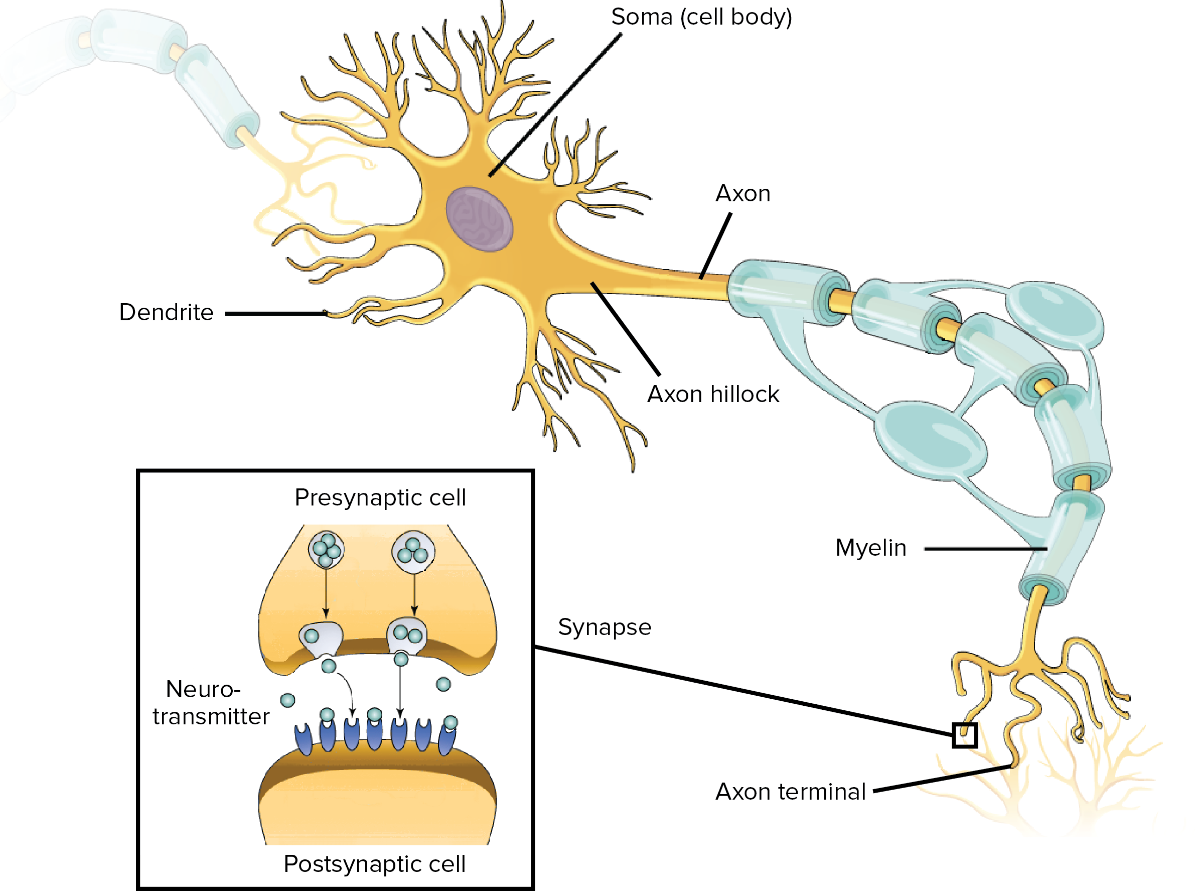
The neuron which receives the neurotransmitters is called the postsynaptic neuron. The end of each neuron has presynaptic endings and vesicles, which are sacks containing neurotransmitters. When a nerve impulse (or action potential) triggers the release of neurotransmitters, these chemicals are then released into the synapse and then is taken ...
' Assuming all of you deep learning aficionados know the basic structure of an Artificial Neural Network (ANN) and how its building block, the neuron, actually function inside of the network, it must be clear to you that each neuron: Receives input(s) from the input layer or neurons from the previous layers in the network ...
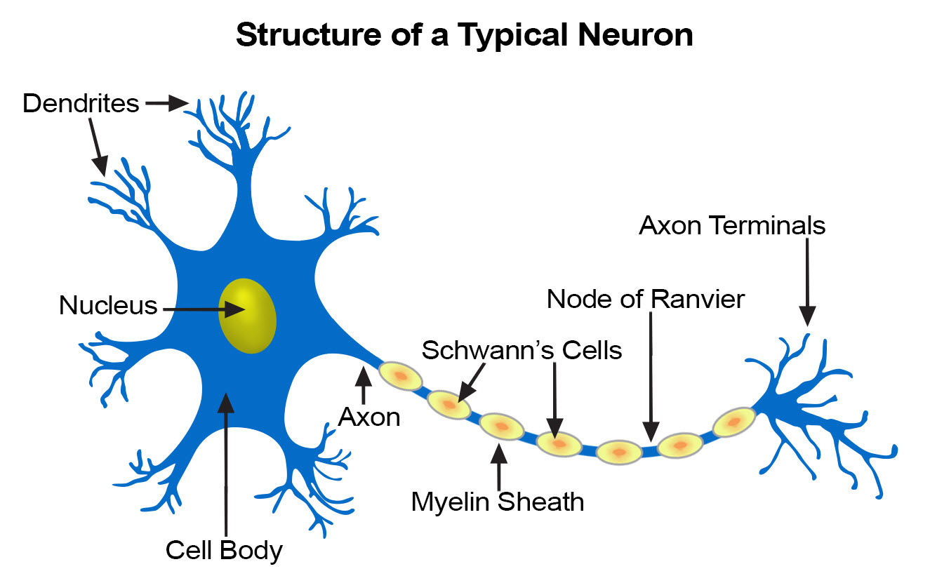
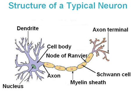
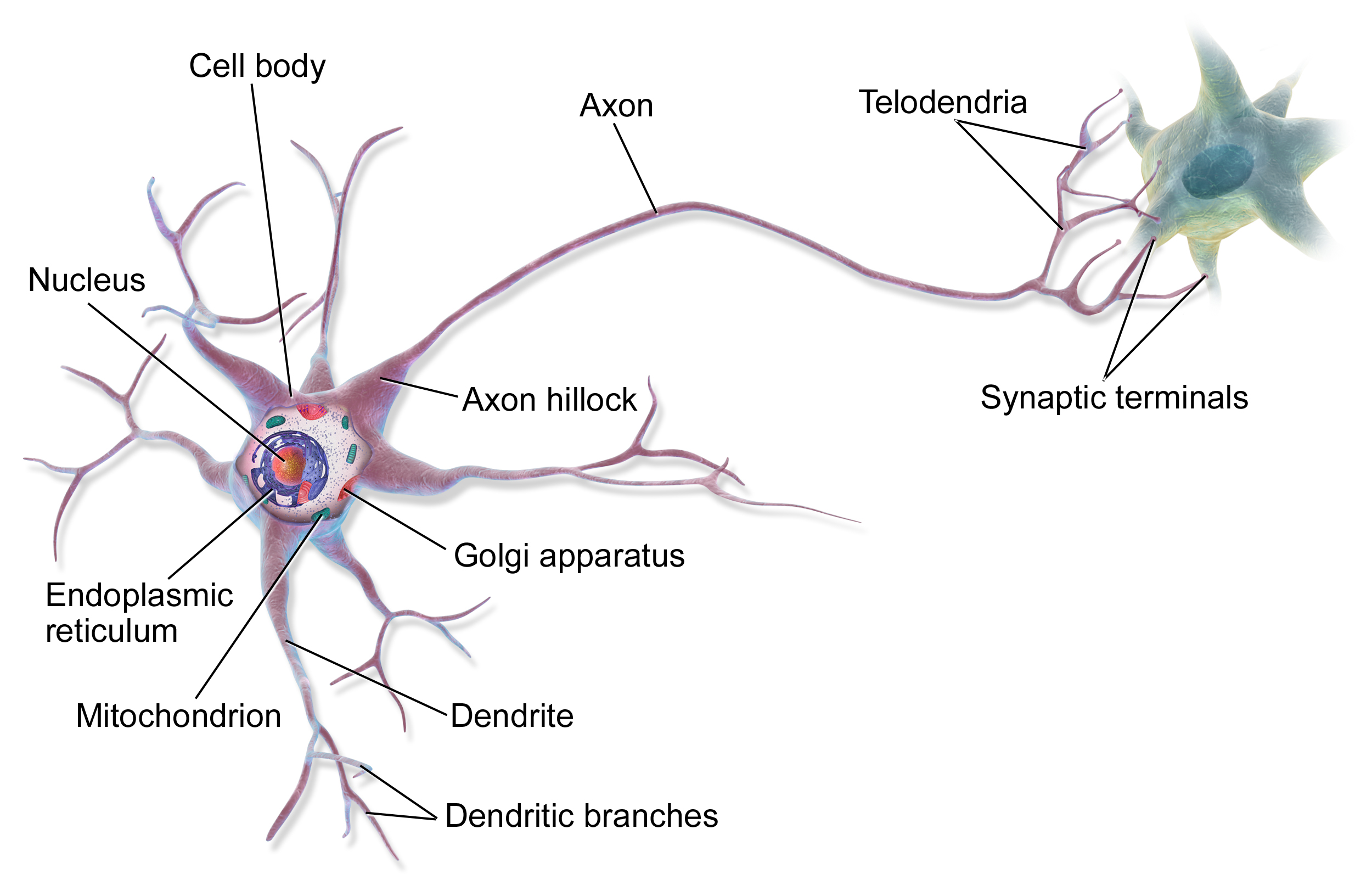
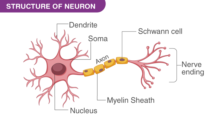
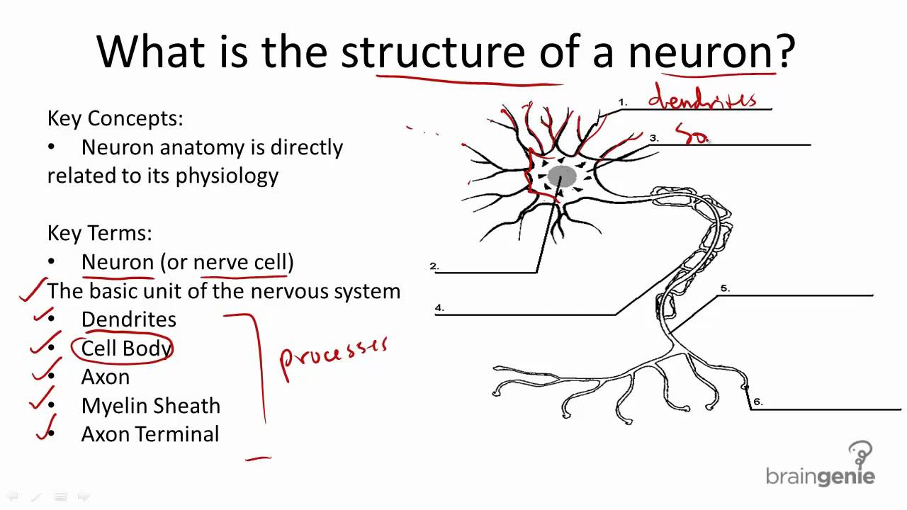
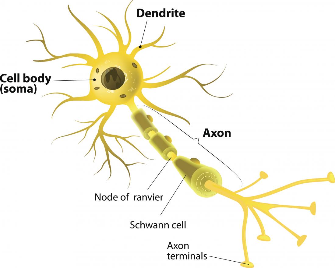
Neuron Facts: 6-10 | Structure of Neurons. 6. There are Neurons of various sizes and shapes. However, a typical Neuron found in a vertebrate body such as humans, the structure of a Neuron can be generally broken down into four broad regions. 7. The four broad regions of a Neuron are: Soma, Dendrites, Axon and Synaptic Terminals.
Neurons are also called nerve cells, and they are specialized cells whose main function is the transmission of various nerve impulses. Their cellular processes include dendrites and axons.
Cell Membrane Neuron Function. The function of neurons is to process and transmit information. The plasma membrane of the neuron is semipermeable, being highly permeable to K+ and slightly permeable to Cl− and Na+. A bacteria diagram clearly enables us to profit extra approximately this single cell organisms which have neither membrane-bounded nucleolus or organelles […]
Neuron diagram and functions It is the coordinating neuron. The neuron s are of three kinds according to their functions. The sensory neuron s conduct nerve currents from the sense-organs to the sensory centres. The motor neuron s which terminate in muscles carry nerve currents from the motor centres to the muscles.
File:neuron.svg - wikimedia commons
Neurons are electrically excitable cells that transmit signals throughout the body. Neurons employ both electrical and chemical components in the transmission of information. Neurons are connected to other neurons at synapses and connected to effector organs or cells at neuroeffector junctions.
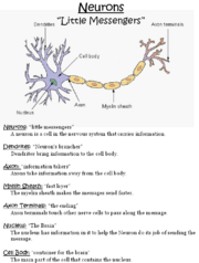
Little messengers - wikieducator
Neuron: Structure & Function- Neuroscience Made Easy- 2 Updated: Nov 29 Neurons are the information processing units of the brain which have a responsibility for sending, receiving, and transmitting electrochemical signals throughout the body .

Neuron+anatomy | ... neuron and their functions (isn't this ...
The axon is a tail-like structure of the neuron which functions in carrying signals away from the soma to the end of the neuron, for the purpose of transmitting signals to other neurons. The dendrites of the neurons are tree-root shaped, located at the end of the neuron.
Overview of neuron structure and function (article) | khan ...
Structure and Function. The neuron's membrane potential gets generated via a difference in the concentration of charged ions. The lipid bilayer of the neuronal cell membrane acts as a capacitor, the transmembrane channels as resistors. This resting (steady-state) potential is critical for the neuron's physiological state, maintained by an ...

Structure and function of neuron
Degradation is when an enzyme changes the structure of the neurotransmitter so that it no longer fits the receptor. Diffusion is when the neurotransmitter begins to diffuse or drift away from the receptor. Reuptake is when the entire neurotransmitter molecule is taken back by the neuron's axon, which initially releases neurotransmitters.
The structure and function of sensory, relay and motor ...
Neurotransmitters are released from one neuron at the presynaptic nerve terminal to cross the synapse where they may be received by the next neuron at a specialized site called a receptor. The major classes of neurotransmitters include amino acids, peptides and monoamines. Here are some of the major neurotransmitters and their primary functions.

File:components of neuron.jpg - wikimedia commons
The Structure and Function of Sensory, Relay and Motor Neurons. The nervous system is composed of specialised cells called neurons. The neurons form pathways in the brain and throughout the body by being connected to one another by synapses. There are about 100 billion neurons or nerve cells in the average nervous system.

Neuron structure and function - lessons - blendspace
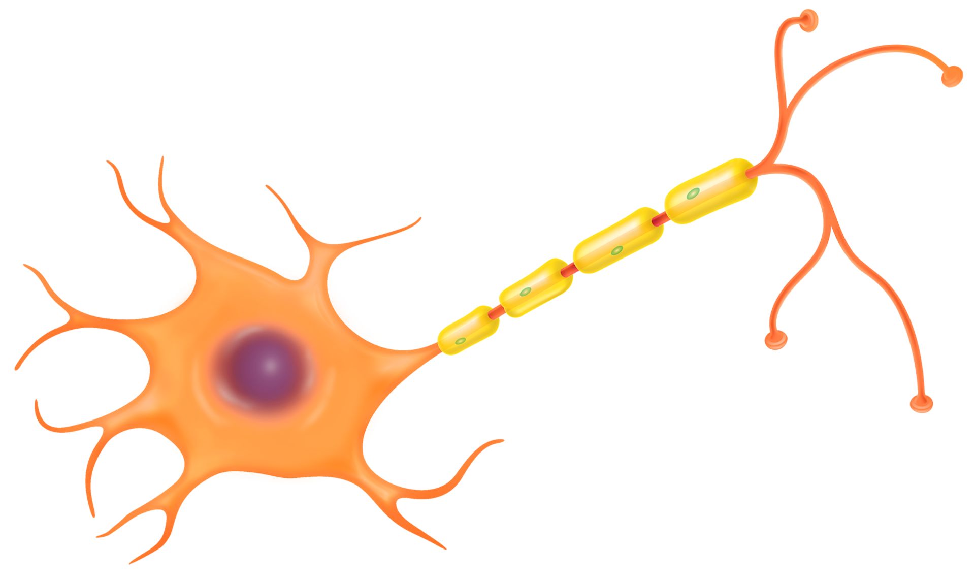
The structure of neurons contains two main components, dendrites and axons, that polarize and depolarize cells. Those two components are responsible for the function of sending signals among cells....
Overview of neuron structure and function (article) | khan ...
Excitatory neurotransmitters function to activate receptors on the postsynaptic membrane and enhance the effects of the action potential, while inhibitory neurotransmitters function to prevent an action potential. In addition to the above classification, neurotransmitters can also be classified based on their chemical structure:
Brain anatomy and how the brain works | johns hopkins medicine
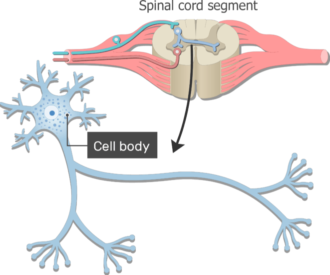
The cell body, also known as the soma, is the part of a neuron that contains the nucleus and controls cell function. Explore the definition, function, and types of cell bodies and learn about neurons.
How does information enter and exit a neuron? how is this ...
Motor neurons are responsible for integrating signals from the brain to the muscles, glands, and organs that intend to carry out the required motor function. Motor neurons allow us to move, talk, eat, swallow, and breathe, therefore without these cells, we would not be able to complete many basic life functions.
Seer training: nerve tissue
Seer training: neurons & glial cells
The neuron
A labelled diagram of neuron with detailed explanations

Neuron structure and function – the alcohol pharmacology ...
Nerve cell function | nerve cell diagram | dk find out
Structure and function of neurons and hebb's theory (2 part ...
Neuron structure and function
Get an overview of neuron cells- cusabio

Structure of neurons: what is a neuron? types, structure ...
Neuron - wikipedia
What is a neuron? write the structure and functions of a ...

Neurons (nerve cells) structure, function & types - simply ...

Structure & function - the nervous system: anatomy and more
Nervous system: structure, function and diagram | kenhub

Basic neuron types. | download scientific diagram
What is a neuron? - definition, structure, parts and function

Neuron structure.
Neurons | boundless psychology

Diagram quiz on neuron structure and function (labeling quiz)
8 1 3 nerve cell structure and function

Nervous system | the partnership in education

Parts and functions of a neuron flashcards | quizlet

Neuron structure & function - ppt download
Neurons: what are they and how do they work?

Structure and functions of neuron - msrblog

Structure of a typical motor neuron dendrites neurofibril ...
Neuron - basic structure and functions
0 Response to "40 neuron diagram and functions"
Post a Comment